Modified Subtraction Coronary CT Angiography with a Two-Breathhold Technique: Image Quality and Diagnostic Accuracy in Patients with Coronary Calcifications
- Affiliations
-
- 1Department of Radiology, Zhongshan Hospital, Fudan University, Shanghai, China. zengmengsu@outlook.com
- 2Shanghai Institute of Medical Imaging, Shanghai, China.
- 3Department of Cardiology, Zhongshan Hospital, Fudan University, Shanghai, China.
- 4Department of Radiology, Tongji Hospital, Tongji Medical College, Huazhong University of Science and Technology, Wuhan, China.
- KMID: 2467025
- DOI: http://doi.org/10.3348/kjr.2018.0845
Abstract
OBJECTIVE
To evaluate a modified subtraction coronary computed tomography angiography (CCTA) technique with a two-breathhold approach in terms of image quality and stenosis grading of calcified coronary segments and in the detection of significant coronary stenosis in segments with severe calcification.
MATERIALS AND METHODS
The institutional board approved this study, and all subjects provided written consent. A total of 128 patients were recruited into this trial, of which 32 underwent subtraction CCTA scans and invasive coronary angiography (ICA). The average Agatston score was 356 ± 145. In severely calcified coronary segments, the presence of significant (> 50%) stenosis was assessed on both conventional CCTA and subtraction CCTA images, and the results were finally compared with ICA findings as the gold standard.
RESULTS
For severely calcified segments, the image quality in conventional CCTA significantly improved from 2.51 ± 0.98 to 3.12 ± 0.94 in subtraction CCTA (p < 0.001). In target segments, specificity (70% vs. 87%; p = 0.005) and positive predictive value (61% vs. 79%, p < 0.01) were improved using subtraction CCTA in comparison with conventional CCTA, with no loss in the negative predictive value. The segment-based diagnostic accuracy for detecting significant stenosis was significantly better in subtraction CCTA than in conventional CCTA (area under the receiver operating characteristic curve, 0.94 vs. 0.85; p = 0.03).
CONCLUSION
This modified subtraction CCTA method showed lower misregistration and better image quality in patients with limited breathhold capability. In comparison with conventional CCTA, modified subtraction CCTA would allow stenosis regrading and improve the diagnostic accuracy in coronary segments with severe calcification.
MeSH Terms
Figure
-

Fig. 1 Scan protocol for imaging acquisition for modified subtraction CCTA.BH time is interval between end of BH announcement to end of contrast scan, and also to end of mask scan respectively. BH = breath-holding, CCTA = coronary computed tomography angiography
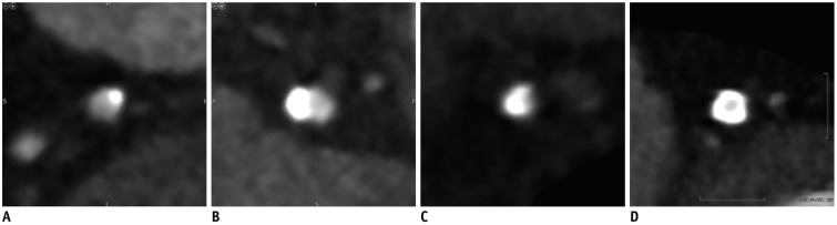
Fig. 2 Examples of coronary atherosclerotic plaque calcification by CT images.A. Mildly calcified (cross-sectional arc calcium < 90°). B. Moderately calcified (cross-sectional arc calcium 90°–180°). C, D. Severely calcified (cross-sectional arc calcium > 180°) segments. Segment calcification was measured by using cross-sectional arc method.
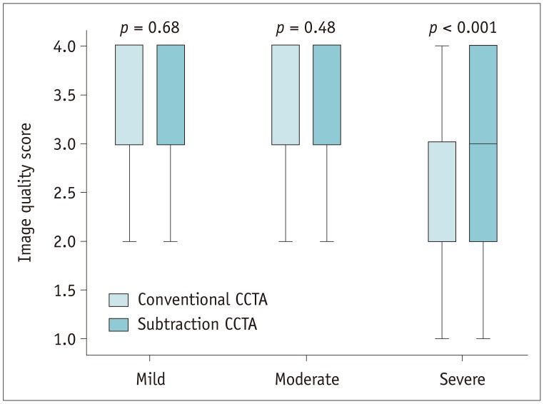
Fig. 3 Image quality of conventional and subtraction computed tomography angiography in different degrees of segment-based plaque calcifications.Image quality in subtraction CCTA is significantly improved in segments with severe calcification (2.51 ± 0.98 to 3.12 ± 0.94, p < 0.001). However, image quality is only slightly improved in segments with mild calcification (3.68 ± 0.54 to 3.71 ± 0.49) and segments with moderate calcification (3.49 ± 0.65 to 3.56 ± 0.59), but not significantly (p = 0.68 and p = 0.48, respectively). Image quality score: 1 = uninterpretable, 2 = poor image quality, 3 = adequate image quality, 4 = good image quality.
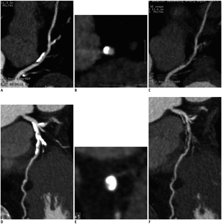
Fig. 4 Case showing stenosis regrading in conventional CCTA and subtraction.A, B. Moderate calcification is observed in left ascending artery on conventional images. C. After performing subtraction, calcification is eliminated. D–F. Degree of stenosis is assessed as mild in both subtraction and conventional CCTA. Corresponding subtraction process for severe plaque calcification at left anterior descending coronary artery from another patient. Moderate stenosis can be seen in subtraction CCTA, while severe stenosis is rated in conventional CCTA.
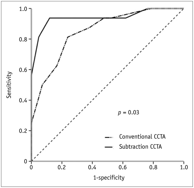
Fig. 5 Area under receiver operating characteristic curves of conventional CCTA and subtraction CCTA versus invasive coronary angiography: AUC for conventional CCTA is 0.85 (95% CI, 0.74–0.96).AUC for subtraction CCTA is 0.94 (95% CI, 0.86–1.00). ACU = area under curve, CI = confidence interval
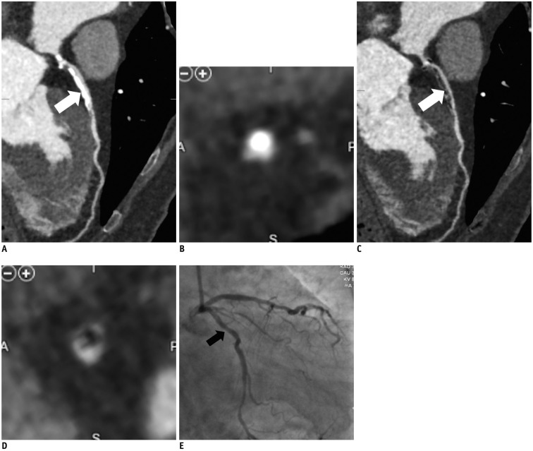
Fig. 6 68-year-old man with suspected coronary artery disease.From CPR image (A) and cross-sectional image (B) in conventional CCTA, severe calcification is observed in proximal portion of left anterior descending artery, which makes it difficult to assess lumen (arrow). In cross-sectional image (C) and CPR cross-sectional image (D) after subtraction, no significant stenosis is depicted (arrow). There is no significant stenosis determined by invasive coronary angiography (arrow) (E). CPR = curved planar reformation
Reference
-
1. Miller JM, Rochitte CE, Dewey M, Arbab-Zadeh A, Niinuma H, Gottlieb I, et al. Diagnostic performance of coronary angiography by 64-row CT. N Engl J Med. 2008; 359:2324–2336. PMID: 19038879.
Article2. Dewey M, Vavere AL, Arbab-Zadeh A, Miller JM, Sara L, Cox C, et al. Patient characteristics as predictors of image quality and diagnostic accuracy of MDCT compared with conventional coronary angiography for detecting coronary artery stenoses: CORE-64 Multicenter International Trial. AJR Am J Roentgenol. 2010; 194:93–102. PMID: 20028910.
Article3. Meijboom WB, Meijs MF, Schuijf JD, Cramer MJ, Mollet NR, van Mieghem CA, et al. Diagnostic accuracy of 64-slice computed tomography coronary angiography: a prospective, multicenter, multivendor study. J Am Coll Cardiol. 2008; 52:2135–2144. PMID: 19095130.4. Zhang S, Levin DC, Halpern EJ, Fischman D, Savage M, Walinsky P. Accuracy of MDCT in assessing the degree of stenosis caused by calcified coronary artery plaques. AJR Am J Roentgenol. 2008; 191:1676–1683. PMID: 19020235.
Article5. Dewey M. Coronary CT versus MR angiography: pro CT--the role of CT angiography. Radiology. 2011; 258:329–339. PMID: 21273517.
Article6. Palumbo AA, Maffei E, Martini C, Tarantini G, Di Tanna GL, Berti E, et al. Coronary calcium score as gatekeeper for 64-slice computed tomography coronary angiography in patients with chest pain: per-segment and per-patient analysis. Eur Radiol. 2009; 19:2127–2135. PMID: 19387651.
Article7. Ahn SJ, Kang DK, Sun JS, Yoon MH. Accuracy and predictive value of coronary computed tomography angiography for the detection of obstructive coronary heart disease in patients with an Agatston calcium score above 400. J Comput Assist Tomogr. 2013; 37:387–394. PMID: 23674010.
Article8. Yoshioka K, Tanaka R, Muranaka K. Subtraction coronary CT angiography for calcified lesions. Cardiol Clin. 2012; 30:93–102. PMID: 22304952.
Article9. Amanuma M, Kondo T, Sano T, Sekine T, Takayanagi T, Matsutani H, et al. Subtraction coronary computed tomography in patients with severe calcification. Int J Cardiovasc Imaging. 2015; 31:1635–1642. PMID: 26288954.
Article10. Yoshioka K, Tanaka R, Muranaka K, Sasaki T, Ueda T, Chiba T, et al. Subtraction coronary CT angiography using second-generation 320-detector row CT. Int J Cardiovasc Imaging. 2015; 31(Suppl 1):51–58. PMID: 25721727.
Article11. Viladés Medel D, Leta R, Alomar Serralach X, Carreras Costa F, Pons-Lladó G. Reliability of a new method for coronary artery calcium or metal subtraction by 320-row cardiac CT. Eur Radiol. 2016; 26:3208–3214. PMID: 26662029.
Article12. Amanuma M, Kondo T, Sano T, Takayanagi T, Matsutani H, Sekine T, et al. Assessment of coronary in-stent restenosis: value of subtraction coronary computed tomography angiography. Int J Cardiovasc Imaging. 2016; 32:661–670. PMID: 26662268.
Article13. Yoshioka K, Tanaka R, Takagi H, Nagata K, Chiba T, Takeda K, et al. Diagnostic accuracy of a modified subtraction coronary CT angiography method with short breath-holding time: a feasibility study. Br J Radiol. 2016; 89:20160489. PMID: 27439592.
Article14. Fuchs A, Kühl JT, Chen MY, Helqvist S, Razeto M, Arakita K, et al. Feasibility of coronary calcium and stent image subtraction using 320-detector row CT angiography. J Cardiovasc Comput Tomogr. 2015; 9:393–398. PMID: 26091841.
Article15. Fuchs A, Kühl JT, Chen MY, Viladés Medel D, Alomar X, Shanbhag SM, et al. Subtraction CT angiography improves evaluation of significant coronary artery disease in patients with severe calcifications or stents-the C-Sub 320 multicenter trial. Eur Radiol. 2018; 28:4077–4085. PMID: 29696430.
Article16. Agatston AS, Janowitz WR, Hildner FJ, Zusmer NR, Viamonte M Jr, Detrano R. Quantification of coronary artery calcium using ultrafast computed tomography. J Am Coll Cardiol. 1990; 15:827–832. PMID: 2407762.
Article17. Funama Y, Utsunomiya D, Taguchi K, Oda S, Shimonobo T, Yamashita Y. Automatic exposure control at single- and dual-heartbeat CTCA on a 320-MDCT volume scanner: effect of heart rate, exposure phase window setting, and reconstruction algorithm. Phys Med. 2014; 30:385–390. PMID: 24225011.
Article18. Hausleiter J, Meyer T, Hermann F, Hadamitzky M, Krebs M, Gerber TC, et al. Estimated radiation dose associated with cardiac CT angiography. JAMA. 2009; 301:500–507. PMID: 19190314.
Article19. Razeto M, Mohr B, Arakita K, Schuijf JD, Fuchs A, Kühl JT, et al. Accurate, fully-automated registration of coronary arteries for volumetric CT digital subtraction angiography. In : Proceedings of SPIE - The International Society for Optical Engineering; 2014 March 21; San Diego, CA, USA.20. Vavere AL, Arbab-Zadeh A, Rochitte CE, Dewey M, Niinuma H, Gottlieb I, et al. Coronary artery stenoses: accuracy of 64-detector row CT angiography in segments with mild, moderate, or severe calcification--a subanalysis of the CORE-64 trial. Radiology. 2011; 261:100–108. PMID: 21828192.
Article21. Cerci R, Vavere AL, Miller JM, Yoneyama K, Rochitte CE, Dewey M, et al. Patterns of coronary arterial lesion calcification by a novel, cross-sectional CT angiographic assessment. Int J Cardiovasc Imaging. 2013; 29:1619–1627. PMID: 23702949.
Article22. Raff GL, Abidov A, Achenbach S, Berman DS, Boxt LM, Budoff MJ, et al. Society of Cardiovascular Computed Tomography. SCCT guidelines for the interpretation and reporting of coronary computed tomographic angiography. J Cardiovasc Comput Tomogr. 2009; 3:122–136. PMID: 19272853.23. DeLong ER, DeLong DM, Clarke-Pearson DL. Comparing the areas under two or more correlated receiver operating characteristic curves: a nonparametric approach. Biometrics. 1988; 44:837–845. PMID: 3203132.
Article24. Abdulla J, Pedersen KS, Budoff M, Kofoed KF. Influence of coronary calcification on the diagnostic accuracy of 64-slice computed tomography coronary angiography: a systematic review and meta-analysis. Int J Cardiovasc Imaging. 2012; 28:943–953. PMID: 21667273.
Article25. Earls JP, Berman EL, Urban BA, Curry CA, Lane JL, Jennings RS, et al. Prospectively gated transverse coronary CT angiography versus retrospectively gated helical technique: improved image quality and reduced radiation dose. Radiology. 2008; 246:742–753. PMID: 18195386.
Article26. Machida H, Tanaka I, Fukui R, Shen Y, Ishikawa T, Tate E, et al. Current and novel imaging techniques in coronary CT. Radiographics. 2015; 35:991–1010. PMID: 26046942.
Article27. Sun Z, Ng CK, Xu L, Fan Z, Lei J. Coronary CT angiography in heavily calcified coronary arteries: improvement of coronary lumen visualization and coronary stenosis assessment with image postprocessing methods. Medicine (Baltimore). 2015; 94:e2148. PMID: 26632895.28. Li P, Xu L, Yang L, Wang R, Hsieh J, Sun Z, et al. Blooming artifact reduction in coronary artery calcification by a new de-blooming algorithm: initial study. Sci Rep. 2018; 8:6945. PMID: 29720611.
Article29. Nørgaard BL, Gaur S, Leipsic J, Ito H, Miyoshi T, Park SJ, et al. Influence of coronary calcification on the diagnostic performance of CT angiography derived FFR in coronary artery disease: a substudy of the NXT trial. JACC Cardiovasc Imaging. 2015; 8:1045–1055. PMID: 26298072.
- Full Text Links
-
- Actions
-
Cited
- CITED
-
- Close
- Share
-
- Similar articles
-
- Assessment of the Image Quality and Diagnostic Accuracy of Coronary CT Angiography: Effect of Sublingual Administration of Nitroglycerin
- Coronary CT Angiography
- Assessment of Coronary Stenosis Using Coronary CT Angiography in Patients with High Calcium Scores: Current Limitations and Future Perspectives
- The Diagnostic Accuracy of the 64-slice Multi-detector CT Coronary Angiography for the Assessment of Coronary Artery Stenosis in Symptomatic Patients
- Technical Aspect of Coronary CT Angiography: Imaging Tips and Safety Issues
