Reappraising the neurosurgical significance of the pterion location, morphology, and its relationship to optic canal and sphenoid ridge and neurosurgical implications
- Affiliations
-
- 1Department of Anatomy, G.S.L. Medical College, Rajahmundry, India. drvenkateshkamath@gmail.com
- 2Yenepoya Medical College, Mangalore, India.
- KMID: 2466693
- DOI: http://doi.org/10.5115/acb.18.200
Abstract
- Frontolateral craniotomy procedures have advanced from conventional craniotomy to mini-craniotomy, and to contemporary keyhole surgery. In this context, it is important for the neurosurgeon to precisely locate the pterion. The distance of the pterion center from midpoint of zygomatic arch and posterolateral margin of frontozygomatic suture was studied bilaterally in 50 whole adult skulls in Indian ethnic group. The depth of optic canal and sphenoid ridge from the pterion was recorded bilaterally in fifty cut adult skulls and fifteen three-dimensional computed tomography scans. The suture length, thickness, and morphology were studied. The data were analyzed using SPSS software, two-tailed Student's t test, binary logistic regression and receiver operating characteristic curve for sexual dimorphism. The pterion center was located at a mean distance of 37.02 mm above the midpoint of zygomatic arch, 28.20 mm behind the posterolateral margin of frontozygomatic suture, 42.73 mm lateral to the optic canal and 10.59 mm from the sphenoid ridge. The location did not exhibit sexual dimorphism. In 20% cases the pterion center was 40 mm or more above the midpoint of the zygomatic arch and in 5% cases 35 mm or more posterior to the posterolateral margin of frontozygomatic suture. The mean suture length was 10±3 mm. The mean thickness at the center of the pterion was 3.52±1.45 mm. The commonest variety was sphenoparietal followed by frontotemporal, epipteric, and stellate types. A thorough knowledge of these dimensions has innumerable neurosurgical implications in resection of sellar, parasellar, and paraclinoid tumors and circulatory aneurysms.
MeSH Terms
Figure
-

Fig. 1 The technique of marking the center of the pterion externally. CP marks the location of the pterion. Type, sphenoparietal. CP, center of the pterion; F, frontal bone; P, parietal bone; S, sphenoid bone; T, temporal bone.
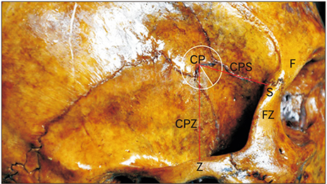
Fig. 2 The location of the pterion as linear distances from specific landmarks. CP, center of the pterion; CPS, distance from the center of the pterion to the posterolateral margin of the frontozygomatic suture; CPZ, distance from the center of the pterion to the midpoint of the zygoma; F, frontal bone; FZ, the frontal process of the zygomatic bone with the fronto-zygomatic suture intervening; S, posterolateral margin of the frontozygomatic suture; Z, midpoint of the zygoma.
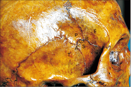
Fig. 3 The technique of measurement of the length of the suture. Type of pterion, sphenoparietal. C, distance between the points A and B and marks the length of the suture; F, frontal bone; P, parietal bone; S, sphenoid bone; T, temporal bone.

Fig. 4 The measurement of depth optic canal and the sphenoid ridge from the pterion. AB is a vertical line drawn at the lateral margin of the optic canal. IP-L is the horizontal distance from the internal location of the pterion to the lateral margin of the optic canal. IP-SR is the linear distance from the internal location of the pterion to the lateral end of the sphenoid ridge. IP, internal location of the pterion; OC, optic canal; SR, a point on the lateral end of the sphenoid ridge.
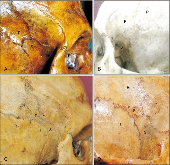
Fig. 5 The four patterns of the pterion as described by Murphy [3]. (A) Sphenoparietal. (B) Frontotemporal. (C) Stellate. (D) Epipteric. E, epipteric bone; F, frontal bone; P, parietal bone; S, sphenoid bone; T, temporal bone.
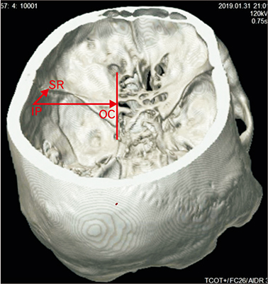
Fig. 6 Measurement of depth optic canal and the sphenoid ridge from the pterion in 3-dimensional computed tomography scan. IP-OC is the horizontal distance from the internal socation of the pterion to the lateral margin of the optic canal. IP-SR is the linear distance from the internal location of the pterion to the lateral end of the sphenoid ridge. IP, internal location of the pterion; OC, optic canal; SR, a point on the lateral end of the sphenoid ridge.
Reference
-
1. Williams PL, Bannister LH, Berry MM, Collins P, Dyson M, Dussek JE, Ferguson MW. Gray's Anatomy: The Anatomical Basis of Medicine and Surgery. 38th ed. London: Churchill Livingstone;1995. p. 583–606.2. Broca P. Instructions craniologiques et craniometriques. Mem Soc Anthrop Paris. 1875; 2:1–203.3. Murphy T. The pterion in the Australian aborigine. Am J Phys Anthropol. 1956; 14:225–244.4. Figueiredo EG, Oliveira AM, Plese JP, Teixeira MJ. Perspective of the frontolateral craniotomies. Arq Neuropsiquiatr. 2010; 68:430–432.5. Figueiredo EG, Deshmukh P, Zabramski JM, Preul MC, Crawford NR, Spetzler RF. The pterional-transsylvian approach: an analytical study. Neurosurgery. 2006; 59:ONS263–ONS269.6. Schick U, Dott U, Hassler W. Surgical management of meningiomas involving the optic nerve sheath. J Neurosurg. 2004; 101:951–959.7. Ngando HM, Maslehaty H, Schreiber L, Blaeser K, Scholz M, Petridis AK. Anatomical configuration of the Sylvian fissure and its influence on outcome after pterional approach for microsurgical aneurysm clipping. Surg Neurol Int. 2013; 4:129.8. Hyun SJ, Hong SC, Kim JS. Side selection of the pterional approach for superiorly projecting anterior communicating artery aneurysms. J Clin Neurosci. 2010; 17:592–596.9. Ma S, Baillie LJ, Stringer MD. Reappraising the surface anatomy of the pterion and its relationship to the middle meningeal artery. Clin Anat. 2012; 25:330–339.10. Ersoy M, Evliyaoglu C, Bozkurt MC, Konuskan B, Tekdemir I, Keskil IS. Epipteric bones in the pterion may be a surgical pitfall. Minim Invasive Neurosurg. 2003; 46:363–365.11. Mwachaka P, Hassanali J, Odula P. Anatomic position of the pterion among Kenyans for lateral skull approaches. Int J Morphol. 2008; 26:931–933.12. Adejuwon SA, Olopade FE, Bolaji M. Study of the location and morphology of the pterion in adult Nigerian skulls. ISRN Anat. 2013; 2013:403937.13. Apinhasmit W, Chompoopong S, Chaisuksunt V, Thiraphatthanavong P, Phasukdee N. Anatomical consideration of pterion and its related references in Thai dry skulls for pterional surgical approach. J Med Assoc Thai. 2011; 94:205–214.14. de Robles P, Fiest KM, Frolkis AD, Pringsheim T, Atta C, St Germaine-Smith C, Day L, Lam D, Jette N. The worldwide incidence and prevalence of primary brain tumors: a systematic review and meta-analysis. Neuro Oncol. 2015; 17:776–783.15. Dandy WE. The practice of surgery. The brain. Hagerstown, MD: W.F. Prior;1932. Vol. 12.16. Cheng WY, Lee HT, Sun MH, Shen CC. A pterion keyhole approach for the treatment of anterior circulation aneurysms. Minim Invasive Neurosurg. 2006; 49:257–262.17. Kamath V, Asif M, Bhat S, Avadhani R. A study on the pterion position variation and its neurosurgical implications. J Anat Soc India. 2016; 65:S33–S39.18. Oguz O, Sanli SG, Bozkir MG, Soames RW. The pterion in Turkish male skulls. Surg Radiol Anat. 2004; 26:220–224.19. Aksu F, Akyer SP, Kale A, Geylan S, Gayretli O. The localization and morphology of pterion in adult West Anatolian skulls. J Craniofac Surg. 2014; 25:1488–1491.20. Ilknur A, Mustafa KI, Sinan B. A comparative study of variation of the pterion of human skulls from 13th and 20th century Anatolia. Int J Morphol. 2009; 27:1291–1298.21. Urzi F, Iannello A, Torrisi A, Foti P, Mortellaro NF, Cavallaro M. Morphological variability of pterion in the human skull. Ital J Anat Embryol. 2003; 108:83–117.22. Aydin ME, Kopuz C, Demir MT, Corumlu U, Kaya AH. Localization of pterion in neonatal cadavers: a morphometric study. Surg Radiol Anat. 2010; 32:545–550.23. Woo EJ, Jung H, Tansatit T. Cranial index in a modern people of Thai ancestry. Anat Cell Biol. 2018; 51:25–30.24. Ashley-Montagu MF. The anthropological significance of the pterion in the primates. Am J Phys Anthropol. 1933; 18:159–336.25. Matsumura G, Kida K, Ichikawa R, Kodama G. Pterion and epipteric bones in Japanese adults and fetuses, with special reference to their formation and variations. Kaibogaku Zasshi. 1991; 66:462–471.26. Hwang K, Kim JH, Baik SH. The thickness of the skull in Korean adults. J Craniofac Surg. 1999; 10:395–399.27. Pensler J, McCarthy JG. The calvarial donor site: an anatomic study in cadavers. Plast Reconstr Surg. 1985; 75:648–651.
- Full Text Links
-
- Actions
-
Cited
- CITED
-
- Close
- Share
-
- Similar articles
-
- Morphological Analysis of the Pterion in Korean
- Incidence and Types of Sphenoethmoid Cell in Patients with Chronic Paranasal Sinusitis
- Optic foramen location on computed tomography
- Pneumatization of the sphenoid sinus and its surrounding neurovascular structures
- Isolated Sphenoid Sinus Mucocele Presenting as Third Nerve Palsy
