Comparison of the Diagnostic Ability of Endoscopic Ultrasonography and Abdominopelvic Computed Tomography in the Diagnosis of Gastric Subepithelial Tumors
- Affiliations
-
- 1Department of Internal Medicine, College of Medicine, Ewha Womans University, Seoul, Korea. shimkn@ewha.ac.kr
- 2Department of Surgery, College of Medicine, Ewha Womans University, Seoul, Korea.
- KMID: 2465799
- DOI: http://doi.org/10.5946/ce.2019.019
Abstract
- BACKGROUND/AIMS
Endoscopic ultrasonography (EUS) is the most efficient imaging modality for gastric subepithelial tumors (SETs). However, abdominopelvic computed tomography (APCT) has other advantages in evaluating the characteristics, local extension, or invasion of SETs to adjacent organs. This study aimed to compare the diagnostic ability of EUS and APCT based on surgical histopathology results.
METHODS
We retrospectively reviewed data from 53 patients who underwent both EUS and APCT before laparoscopic wedge resection for gastric SETs from January 2010 to December 2017 at a single institution. On the basis of histopathology results, we assessed the diagnostic ability of the 2 tests.
RESULTS
The overall accuracy of EUS and APCT was 64.2% and 50.9%, respectively. In particular, the accuracy of EUS vs. APCT for the diagnosis of gastrointestinal stromal tumors (GISTs), leiomyomas, and ectopic pancreas was 83.9% vs. 74.2%, 37.5% vs. 0.0%, and 57.1% vs. 14.3%, respectively. Most of the incorrect diagnoses with EUS involved hypoechoic lesions originating in the fourth echolayer, with the most common misdiagnosed lesions being GISTs mistaken for leiomyomas and vice versa.
CONCLUSIONS
APCT showed a lower overall accuracy than EUS; however, APCT remains a useful modality for malignant/potentially malignant gastric SETs.
Keyword
MeSH Terms
Figure
-
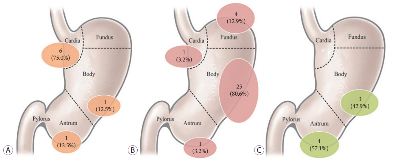
Fig. 1. Distribution of gastric subepithelial tumors: (A) leiomyoma, (B) gastrointestinal stromal tumor, and (C) ectopic pancreas.

Fig. 2. Leiomyoma was misdiagnosed as a gastrointestinal stromal tumor (GIST) based on endoscopic ultrasonography (EUS) findings in a 37-year-old man (Table 4, case 4). (A) Upper endoscopy showed a gastric subepithelial tumor with a size of about 5.0 cm on the cardia. (B) EUS showed a 5.2-cm, well-defined, hypoechoic with a heterogeneous echotexture, and septated lesion mainly originating in the fourth layer. The lesion was presumed to be a GIST. (C) Contrast-enhanced computed tomography axial image showed a 5.1-cm, well-demarcated, and outward-protruding mass with homogeneous enhancement on the cardia. The lesion was presumed to be a GIST. (D) The histopathological result of surgical resection was leiomyoma with a size of 5.2 cm, which was composed of spindle cells as observed on hematoxylin and eosin staining (×10). (E) Immunohistochemical staining was negative for c-kit (×40). (F) Immunohistochemical staining was positive for actin (×40).
Cited by 1 articles
-
Diagnosis of Gastric Subepithelial Tumors Using Endoscopic Ultrasonography or Abdominopelvic Computed Tomography: Which is Better?
Eun Young Park, Gwang Ha Kim
Clin Endosc. 2019;52(6):519-520. doi: 10.5946/ce.2019.188.
Reference
-
1. Hwang JH, Rulyak SD, Kimmey MB. American Gastroenterological Association institute technical review on the management of gastric subepithelial masses. Gastroenterology. 2006; 130:2217–2228.
Article2. Humphris JL, Jones DB. Subepithelial mass lesions in the upper gastrointestinal tract. J Gastroenterol Hepatol. 2008; 23:556–566.
Article3. Hedenbro JL, Ekelund M, Wetterberg P. Endoscopic diagnosis of submucosal gastric lesions. The results after routine endoscopy. Surg Endosc. 1991; 5:20–23.4. Lee DH, Ko YT. Gastric lesions: evaluation with three-dimensional images using helical CT. AJR Am J Roentgenol. 1997; 169:787–789.
Article5. Kim SY, Kim KO. Management of gastric subepithelial tumors: the role of endoscopy. World J Gastrointest Endosc. 2016; 8:418–424.
Article6. Sakamoto H, Kitano M, Kudo M. Diagnosis of subepithelial tumors in the upper gastrointestinal tract by endoscopic ultrasonography. World J Radiol. 2010; 2:289–297.
Article7. Chak A. EUS in submucosal tumors. Gastrointest Endosc. 2002; 56(4 Suppl):S43–S48.
Article8. Kim JH, Eun HW, Goo DE, Shim CS, Auh YH. Imaging of various gastric lesions with 2D MPR and CT gastrography performed with multidetector CT. Radiographics. 2006; 26:1101–1116. discussion 1117-1118.
Article9. Ra JC, Lee ES, Lee JB, et al. Diagnostic performance of stomach CT compared with endoscopic ultrasonography in diagnosing gastric subepithelial tumors. Abdom Radiol (NY). 2017; 42:442–450.
Article10. Lee CM, Chen HC, Leung TK, Chen YY. Gastrointestinal stromal tumor: computed tomographic features. World J Gastroenterol. 2004; 10:2417–2418.
Article11. Kim JY, Lee JM, Kim KW, et al. Ectopic pancreas: CT findings with emphasis on differentiation from small gastrointestinal stromal tumor and leiomyoma. Radiology. 2009; 252:92–100.
Article12. Megibow AJ, Balthazar EJ, Hulnick DH, Naidich DP, Bosniak MA. CT evaluation of gastrointestinal leiomyomas and leiomyosarcomas. AJR Am J Roentgenol. 1985; 144:727–731.
Article13. Goto O, Kambe H, Niimi K, et al. Discrepancy in diagnosis of gastric submucosal tumor among esophagogastroduodenoscopy, CT, and endoscopic ultrasonography: a retrospective analysis of 93 consecutive cases. Abdom Imaging. 2012; 37:1074–1078.
Article14. Okten RS, Kacar S, Kucukay F, Sasmaz N, Cumhur T. Gastric subepithelial masses: evaluation of multidetector CT (multiplanar reconstruction and virtual gastroscopy) versus endoscopic ultrasonography. Abdom Imaging. 2012; 37:519–530.
Article15. Lim TW, Choi CW, Kang DH, Kim HW, Park SB, Kim SJ. Endoscopic ultrasound without tissue acquisition has poor accuracy for diagnosing gastric subepithelial tumors. Medicine (Baltimore). 2016; 95:e5246.
Article16. Moon JS. Role of endoscopic ultrasonography in guidingtreatment plans for upper gastrointestinal subepithelial tumors. Clin Endosc. 2016; 49:220–225.17. Kang JH, Lim JS, Kim JH, et al. Role of EUS and MDCT in the diagnosis of gastric submucosal tumors according to the revised pathologic concept of gastrointestinal stromal tumors. Eur Radiol. 2009; 19:924–934.
Article18. Ludwig DJ, Traverso LW. Gut stromal tumors and their clinical behavior. Am J Surg. 1997; 173:390–394.
Article19. Caletti G, Fusaroli P, Togliani T, Bocus P, Roda E. Endosonography in gastric lymphoma and large gastric folds. Eur J Ultrasound. 2000; 11:31–40.
Article20. Schizas D, Ntanasis-Stathopoulos I, Tsilimigras DI, et al. The role of endoscopic ultrasound in the diagnosis and management of primary gastric lymphoma. Gastroenterol Res Pract. 2017; 2017:2397430.
Article21. Gossios K, Katsimbri P, Tsianos E. CT features of gastric lymphoma. Eur Radiol. 2000; 10:425–430.
Article22. Ghai S, Pattison J, Ghai S, O’Malley ME, Khalili K, Stephens M. Primary gastrointestinal lymphoma: spectrum of imaging findings with pathologic correlation. Radiographics. 2007; 27:1371–1388.
Article23. Lin YM, Chiu NC, Li AF, Liu CA, Chou YH, Chiou YY. Unusual gastric tumors and tumor-like lesions: radiological with pathological correlation and literature review. World J Gastroenterol. 2017; 23:2493–2504.
Article24. Cho JS, Shin KS, Kwon ST, et al. Heterotopic pancreas in the stomach: CT findings. Radiology. 2000; 217:139–144.
Article25. Lee MJ, Lim JS, Kwon JE, et al. Gastric true leiomyoma: computed tomographic findings and pathological correlation. J Comput Assist Tomogr. 2007; 31:204–208.26. Taylor AJ, Stewart ET, Dodds WJ. Gastrointestinal lipomas: a radiologic and pathologic review. AJR Am J Roentgenol. 1990; 155:1205–1210.
Article27. Maeda H, Okabayashi T, Nishimori I, et al. Diagnostic challenge to distinguish gastric duplication cyst from pancreatic cystic lesions in adult. Intern Med. 2007; 46:1101–1104.
Article28. Shin SY, Lee SJ, Jun JH, et al. Mucosal incision and forceps biopsy for reliable tissue sampling of gastric subepithelial tumors. Clin Endosc. 2017; 50:64–68.
Article29. Lee JS, Kim JJ, Park SM. Laparoscopic gastric wedge resection and prophylactic antireflux surgery for a submucosal tumor of gastroesophageal junction. J Gastric Cancer. 2011; 11:131–134.
Article30. Seo SW, Hong SJ, Han JP, et al. Accuracy of a scoring system for the differential diagnosis of common gastric subepithelial tumors based on endoscopic ultrasonography. J Dig Dis. 2013; 14:647–653.
Article31. Kim DJ, Lee JH, Kim W. Laparoscopic resection for 125 gastroduodenal submucosal tumors. Ann Surg Treat Res. 2014; 86:199–205.
Article32. Lee HH, Hur H, Jung H, Jeon HM, Park CH, Song KY. Analysis of 151 consecutive gastric submucosal tumors according to tumor location. J Surg Oncol. 2011; 104:72–75.
Article
- Full Text Links
-
- Actions
-
Cited
- CITED
-
- Close
- Share
-
- Similar articles
-
- Diagnosis of Gastric Subepithelial Tumors Using Endoscopic Ultrasonography or Abdominopelvic Computed Tomography: Which is Better?
- Endoscopic Ultrasonography in the Diagnosis of Gastric Subepithelial Lesions
- Gastric Subepithelial Tumor Diagnosed by Transabdominal Ultrasonography
- Incidental Gastrointestinal Subepithelial Mass
- Common Gastric Subepithelial Tumors in Koreans
