Intractable Headache Related to Intraventricular Glioblastoma: A Case Report and Literature Review
- Affiliations
-
- 1Department of Medicine, Graduate School, Kyung Hee University, Seoul, Korea.
- 2Department of Radiology, Kyung Hee University Hospital, Kyung Hee University College of Medicine, Seoul, Korea. bandilee@khu.ac.kr
- KMID: 2464921
- DOI: http://doi.org/10.3348/jksr.2019.80.6.1241
Abstract
- Although various neoplasms may develop in the ventricular system, glioblastomas are rare. An 80-year-old woman visited our hospital with intractable headache related to a right ventricular large mass, which exhibited heterogeneous enhancement involving the body, trigone, and occipital horn of the right lateral ventricle on MRI. The mass was diagnosed as glioblastoma multiforme on surgical pathology. Herein, the authors present a case and review the existing literature with regarding to incidence, pathophysiology, prognostic factors, imaging and pathologic findings of intraventricular glioblastoma multiforme.
MeSH Terms
Figure
-
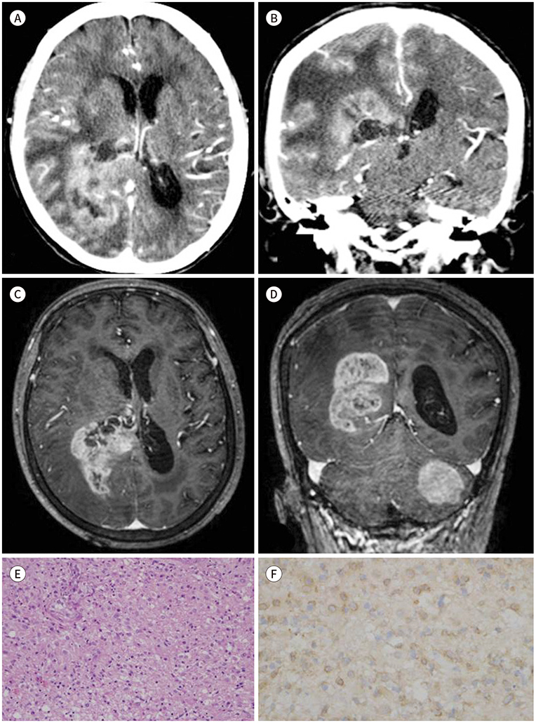
Fig. 1 Intraventricular glioblastoma multiforme in an 80-year-old woman, presenting with intractable headache. A, B. CT images showing a multilobulated, large mass with heterogeneous enhancement in the right parietooccipital area. The mass extends downward along the body, trigone, and occipital horn of the ipsilateral lateral ventricle on axial (A) and coronal (B) images. C, D. Axial (C) and coronal (D) MR images reveal that the heterogeneously enhancing mass is located from the body to occipital horn of right lateral ventricle. A tentorial meningioma is found by chance (D). E. Histopathology shows nuclear polymorphism with several multinucleated giant cells and high degree of cellularity (hematoxylin-eosin staining, × 200). F. Immunohistochemical staining of the histopathology shows positive results for glial fibrillary acidic protein (× 200).
Reference
-
1. Kim YJ, Lee SK, Cho MK, Kim YJ. Intraventricular glioblastoma multiforme with previous history of intracerebral hemorrhage : a case report. J Korean Neurosurg Soc. 2008; 44:405–408.2. Salcman M. Glioblastoma and malignant astrocytoma. In : Kaye A, Laws E, editors. Brain tumors: an encyclopedic approach. Edinburgh: Churchill Livingstone;1995. p. 449–477.3. Park P, Choksi VR, Gala VC, Kaza AR, Murphy HS, Ramnath S. Well-circumscribed, minimally enhancing glioblastoma multiforme of the trigone: a case report and review of the literature. AJNR Am J Neuroradiol. 2005; 26:1475–1478.4. Sarsilmaz A, Gelal F, Apaydin M, Varer M, Bezircioglu H, Rezanko T. Intraventricular glioblastoma multiforme: a pediatric case report. J Pediatr Hematol Oncol. 2010; 32:519–522.5. Jeon YK, Park K, Park CK, Paek SH, Jung HW, Park SH. Chromosome 1p and 19q status and p53 and p16 expression patterns as prognostic indicators of oligodendroglial tumors: a clinicopathological study using fluorescence in situ hybridization. Neuropathology. 2007; 27:10–20.6. Ben Nsir A, Gdoura Y, Thai QA, Zhani Kassar A, Hattab N, Jemel H. Intraventricular glioblastomas. World Neurosurg. 2016; 88:126–131.7. Doetsch F, Caillé I, Lim DA, García-Verdugo JM, Alvarez-Buylla A. Subventricular zone astrocytes are neural stem cells in the adult mammalian brain. Cell. 1999; 97:703–716.8. Quiñones-Hinojosa A, Sanai N, Soriano-Navarro M, Gonzalez-Perez O, Mirzadeh Z, Gil-Perotin S, et al. Cellular composition and cytoarchitecture of the adult human subventricular zone: a niche of neural stem cells. J Comp Neurol. 2006; 494:415–434.9. Quiñones-Hinojosa A, Chaichana K. The human subventricular zone: a source of new cells and a potential source of brain tumors. Exp Neurol. 2007; 205:313–324.10. Mandour C, El Mostarchid B. Intra ventricular glioblastoma. Pan Afr Med J. 2014; 18:100.
- Full Text Links
-
- Actions
-
Cited
- CITED
-
- Close
- Share
-
- Similar articles
-
- Glioblastoma in a Patient with Neurofibromatosis Type 1: A Case Report and Review of the Literature
- Intraventricular Glioblastoma Multiforme with Previous History of Intracerebral Hemorrhage : A Case Report
- Two Cases of Intraventricular Arachnoid Cyst
- Continuous Intraventricular Morphine Infusion for Control of Pain in Terminal Cancer Patients
- Cerebellar Glioblastoma Presenting as a Cerebellar Hemorrhage in a Child
