Three-dimensional analysis of tooth movement in Class II malocclusion treatment using arch wire with continuous tip-back bends and intermaxillary elastics
- Affiliations
-
- 1Department of Orthodontics, School of Dentistry, Wonkwang University, Iksan, Korea. sangkim@wku.ac.kr
- KMID: 2464198
- DOI: http://doi.org/10.4041/kjod.2019.49.6.349
Abstract
OBJECTIVE
The aim of this study was to analyze three-dimensional (3D) changes in maxillary dentition in Class II malocclusion treatment using arch wire with continuous tip-back bends or compensating curve, together with intermaxillary elastics by superimposing 3D virtual models.
METHODS
The subjects were 20 patients (2 men and 18 women; mean age 20 years 7 months ± 3 years 9 months) with Class II malocclusion treated using 0.016 × 0.022-inch multiloop edgewise arch wire with continuous tip-back bends or titanium molybdenum alloy ideal arch wire with compensating curve, together with intermaxillary elastics. Linear and angular measurements were performed to investigate maxillary teeth displacement by superimposing pre- and post-treatment 3D virtual models using Rapidform 2006 and analyzing the results using paired t-tests.
RESULTS
There were posterior displacement of maxillary teeth (p < 0.01) with distal crown tipping of canine, second premolar and first molar (p < 0.05), expansion of maxillary arch (p < 0.05) with buccoversion of second premolar and first molar (p < 0.01), and distal-in rotation of first molar (p < 0.01). Reduced angular difference between anterior and posterior occlusal planes (p < 0.001), with extrusion of anterior teeth (p < 0.05) and intrusion of second premolar and first molar (p < 0.001) was observed.
CONCLUSIONS
Class II treatment using an arch wire with continuous tip-back bends or a compensating curve, together with intermaxillary elastics, could retract and expand maxillary dentition, and reduce occlusal curvature. These results will help clinicians in understanding the mechanism of this Class II treatment.
MeSH Terms
Figure
-
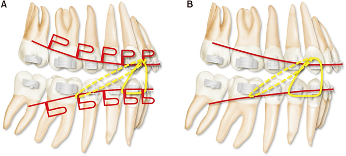
Figure 1 A, 0.016 × 0.022-inch (in) multiloop edgewise arch wire with continuous tipback bends; B, 0.016 × 0.022-in titanium molybdenum alloy ideal arch wire with compensating curve. Intermaxillary elastics (anterior vertical elastics; 3/16 in, 6.5 ounce [oz] and occasionally Class II elastics; 5/16 in, 6.5 oz) were used with these arch wires after alignment and leveling.
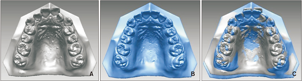
Figure 2 Three-dimensional virtual models. A, Pre-treatment; B, Post-treatment; C, Superimposition using the third medial rugae and the midline raphe as the reference area.
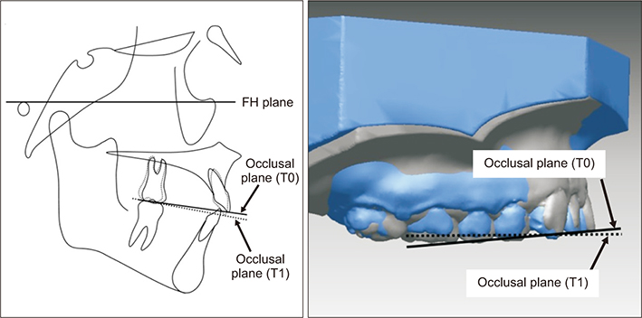
Figure 3 Angles between the maxillary occlusal plane and the Frankfort horizontal (FH) plane at pre-treatment (T0) and post-treatment (T1) were measured on lateral cephalometric radiographs. The angular difference between the occlusal planes at T0 and T1 of the three-dimensional (3D) virtual models was compared with that of the lateral cephalometric radiographs. If the angular difference between the 3D virtual model and the lateral cephalometric radiograph was greater than 5°, superposition of the 3D virtual models was reattempted to correct the error.18
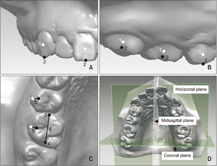
Figure 4 Reference points, lines, and planes. A, Frontal view; B, Sagittal view; C, Occlusal view; D, Reference planes. 1, Facial axis (FA) point; 2, occlusal reference point: the point on the most occlusal side of the FA of the clinical crown; 3, mesial occlusal point; 4, distal occlusal point; 5, FA line: line between the FA point and occlusal reference point; 6, occlusal line: the line between mesiodistal occlusal points.
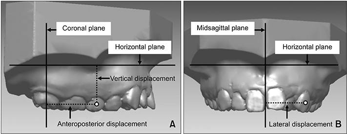
Figure 5 Linear measurements (mm). A, Anteroposterior displacement: the distance from the facial axis (FA) point to the coronal plane. When the tooth was located posterior to the coronal plane, the distance was taken to be negative. For the difference between pre- and post-treatment (T0–T1), positive means posterior movement and negative means anterior movement. Vertical displacement is the distance from the FA point to the horizontal plane. For T0–T1, positive means intrusion and negative means extrusion; B, Lateral displacement: the distance from the FA point to the midsagittal plane. For T0–T1, positive means medial movement and negative means lateral movement.
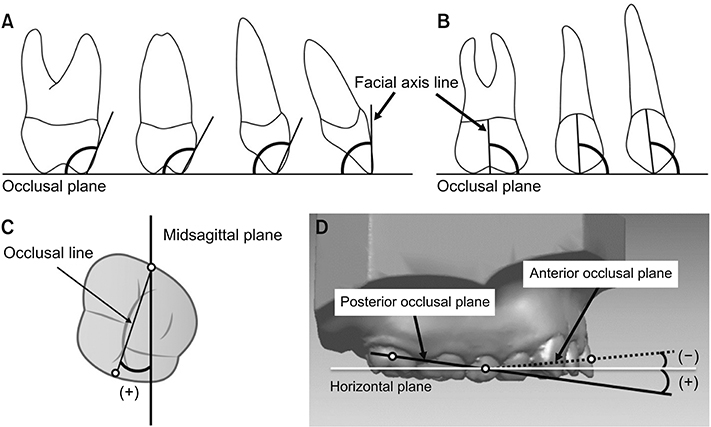
Figure 6 Angular measurements (°). A, Inclination. For the difference between pre- and post-treatment (T0–T1), positive means labioversion or buccoversion and negative means palatoversion; B, Angulation. For T0–T1, positive means distal crown tipping and negative means mesial crown tipping; C, Rotation. When the occlusal line was inclined inward and backward to the midsagittal plane, the angle of rotation was taken to be negative. For T0–T1, positive means distal-in rotation and negative means mesial-in rotation; D, Angles of anterior occlusal plane (AOP) and posterior occlusal plane (POP). When the AOP or POP was inclined forward and upward with respect to the horizontal plane, the angle of AOP or POP was taken to be negative. For T0–T1, positive means counterclockwise rotation and negative means clockwise rotation.
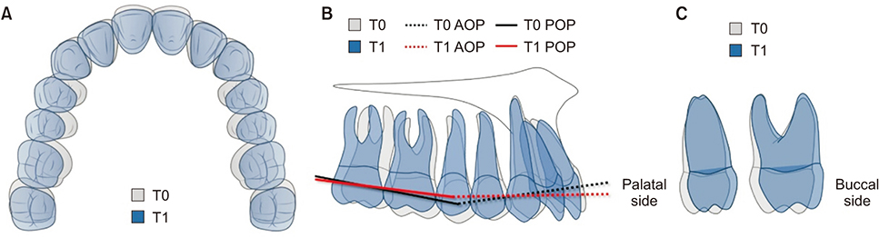
Figure 7 Schematic drawing of treatment results using arch wire with continuous tip-back bends or a compensating curve, together with intermaxillary elastics. We observed posterior displacement of the maxillary teeth and expansion of the maxillary arch with distal-in rotation of the first molar (A). There were distal crown tipping of the canine, second premolar, and first molar (B); and buccoversion of the second premolar and first molar (C). Reduced occlusal curvature was observed as a result of extrusion of the incisor and canine and intrusion of the second premolar and first molar (B). T0, Pre-treatment; T1, post-treatment; AOP, anterior occlusal plane; POP, posterior occlusal plane.
Reference
-
1. Kim YH. Anterior openbite and its treatment with multiloop edgewise archwire. Angle Orthod. 1987; 57:290–321.2. Liu J, Zou L, Zhao ZH, Welburn N, Yang P, Tang T, et al. Successful treatment of postpeak stage patients with class II division 1 malocclusion using non-extraction and multiloop edgewise archwire therapy: a report on 16 cases. Int J Oral Sci. 2009; 1:207–216.
Article3. Locatelli R, Bednar J, Dietz VS, Gianelly AA. Molar distalization with superelastic NiTi wire. J Clin Orthod. 1992; 26:277–279.4. Carano A, Testa M. The distal jet for upper molar distalization. J Clin Orthod. 1996; 30:374–380.5. Keles A. Maxillary unilateral molar distalization with sliding mechanics: a preliminary investigation. Eur J Orthod. 2001; 23:507–515.
Article6. Duran GS, Görgülü S, Dindaroğlu F. Three-dimensional analysis of tooth movements after palatal miniscrew-supported molar distalization. Am J Orthod Dentofacial Orthop. 2016; 150:188–197.
Article7. Ali D, Mohammed H, Koo SH, Kang KH, Kim SC. Three-dimensional evaluation of tooth movement in Class II malocclusions treated without extraction by orthodontic mini-implant anchorage. Korean J Orthod. 2016; 46:280–289.
Article8. Chang YI, Shin SJ, Baek SH. Three-dimensional finite element analysis in distal en masse movement of the maxillary dentition with the multiloop edgewise archwire. Eur J Orthod. 2004; 26:339–345.
Article9. Enacar A, Ugur T, Toroglu S. A method for correction of open bite. J Clin Orthod. 1996; 30:43–48.10. Küçükkeleş N, Acar A, Demirkaya AA, Evrenol B, Enacar A. Cephalometric evaluation of open bite treatment with NiTi arch wires and anterior elastics. Am J Orthod Dentofacial Orthop. 1999; 116:555–562.
Article11. Min S, Chung C, Hwang CJ, Cha JY. Non-extraction treatment in Class III malocclusion by using improved superelastic NiTi wire. Korean J Orthod. 2011; 41:297–306.
Article12. Shin SJ, Chang YI. Three dimensional finite element analysis of the phenomenon during distal en masse movement of the maxillary dentition. Korean J Orthod. 1998; 28:563–580.13. Malkoc S, Sari Z, Usumez S, Koyuturk AE. The effect of head rotation on cephalometric radiographs. Eur J Orthod. 2005; 27:315–321.
Article14. Gaddam R, Shashikumar HC, Lokesh NK, Suma T, Arya S, Shwetha GS. Assessment of image distortion from head rotation in lateral cephalometry. J Int Oral Health. 2015; 7:35–40.15. Trpkova B, Major P, Prasad N, Nebbe B. Cephalometric landmarks identification and reproducibility: a meta analysis. Am J Orthod Dentofacial Orthop. 1997; 112:165–170.
Article16. Cha BK, Lee JY, Jost-Brinkmann PG, Yoshida N. Analysis of tooth movement in extraction cases using three-dimensional reverse engineering technology. Eur J Orthod. 2007; 29:325–331.
Article17. Choi DS, Jeong YM, Jang I, Jost-Brinkmann PG, Cha BK. Accuracy and reliability of palatal superimposition of three-dimensional digital models. Angle Orthod. 2010; 80:497–503.
Article18. Cho MY, Choi JH, Lee SP, Baek SH. Three-dimensional analysis of the tooth movement and arch dimension changes in Class I malocclusions treated with first premolar extractions: a guideline for virtual treatment planning. Am J Orthod Dentofacial Orthop. 2010; 138:747–757.
Article19. Andrews LF. The six keys to normal occlusion. Am J Orthod. 1972; 62:296–309.
Article20. Hoggan BR, Sadowsky C. The use of palatal rugae for the assessment of anteroposterior tooth movements. Am J Orthod Dentofacial Orthop. 2001; 119:482–488.
Article21. Chen G, Chen S, Zhang XY, Jiang RP, Liu Y, Shi FH, et al. Stable region for maxillary dental cast superimposition in adults, studied with the aid of stable miniscrews. Orthod Craniofac Res. 2011; 14:70–79.
Article22. Joffe L. OrthoCAD: digital models for a digital era. J Orthod. 2004; 31:344–347.23. Yang WS, Kim BH, Kim YH. A study of the regional load deflection rate of multiloop edgewise arch wire. Angle Orthod. 2001; 71:103–109.24. Fushima K, Kitamura Y, Mita H, Sato S, Suzuki Y, Kim YH. Significance of the cant of the posterior occlusal plane in class II division 1 malocclusions. Eur J Orthod. 1996; 18:27–40.
Article25. Park HM, Kim BH, Yang IH, Baek SH. Preliminary three-dimensional analysis of tooth movement and arch dimension change of the maxillary dentition in Class II division 1 malocclusion treated with first premolar extraction: conventional anchorage vs. mini-implant anchorage. Korean J Orthod. 2012; 42:280–290.
Article
- Full Text Links
-
- Actions
-
Cited
- CITED
-
- Close
- Share
-
- Similar articles
-
- Three dimensional finite element analysis of the phenomenon during distal en masse movement of the maxillary dentition
- Non-extraction treatment in Class III malocclusion by using improved superelastic NiTi wire
- A photoelastic study of the effect of loops in arch wire and orthodontic elastics in relief of Curve of Spee
- A photoelastic study of the stress distribution by multiloop edgewise arch wire
- Mechanical analysis on the shape-memory arch wire
