Computer-aided design and manufacturing-based full mouth rehabilitation for a patient with excessive attrition and restricted vertical dimension: A case report
- Affiliations
-
- 1Department of Prosthodontics, School of Dentistry and Dental Research Institute, Seoul National University, Seoul, Korea. proshan@snu.ac.kr
- KMID: 2461159
- DOI: http://doi.org/10.4047/jkap.2019.57.4.495
Abstract
- This study reported the treatment of a patient with excessive worn dentition and limited maxillo-mandibular space for restoration, utilizing the computer-aided design and computer-aided manufacturing (CAD/CAM) technology. After the thorough examination of the patient's occlusal vertical dimension (OVD), full mouth rehabilitation was planned with increase of the OVD. The patient was satisfied with the provisional restorations establishing the increased OVD. The horizontal and vertical data of the patient's jaw relation that the provisional restorations contained were transferred to the definitive metal ceramic fixed prostheses by double scanning and three-dimensional printing. After the fixed restorations were cemented to the abutments, electronic surveying and three-dimensional printing were used to fabricate metal frameworks for the patient's removable partial dentures. The mandibular definitive removable prostheses were delivered to the patient's mouth and the full mouth rehabilitation procedures were completed. The digital technologies used for this case produced fixed and removable restorations satisfactory in masticatory, phonetic and aesthetic functions to both the patient and the dental clinician.
Keyword
MeSH Terms
Figure
-
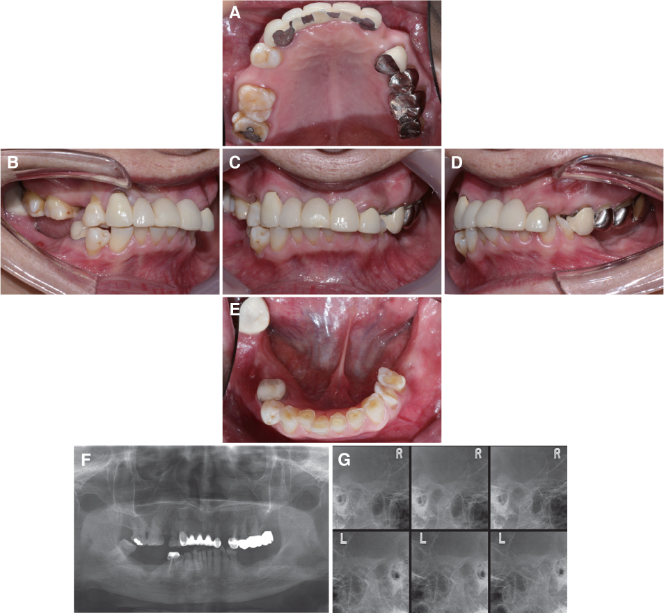
Fig. 1 Preoperative intraoral photographs and radiographic evaluation. (A) Maxillary occlusal view, (B) Right lateral view, (C) Frontal view, (D) Left lateral view, (E) Mandibular occlusal view, (F) Panoramic radiograph, (G) Transcranial view are shown.
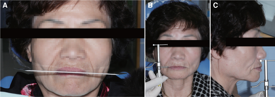
Fig. 2 Preoperative extraoral photos. (A) Occlusal plane analysis, (B, C) Vertical dimension analysis with Willis method are shown.
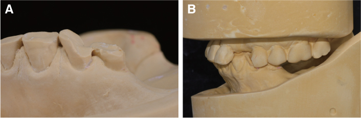
Fig. 3 Diagnostic cast analysis. (A) Excessive worn of mandibular left premolars, (B) Limited maxillo-mandibular space for restoration of left canine and premolars are shown.
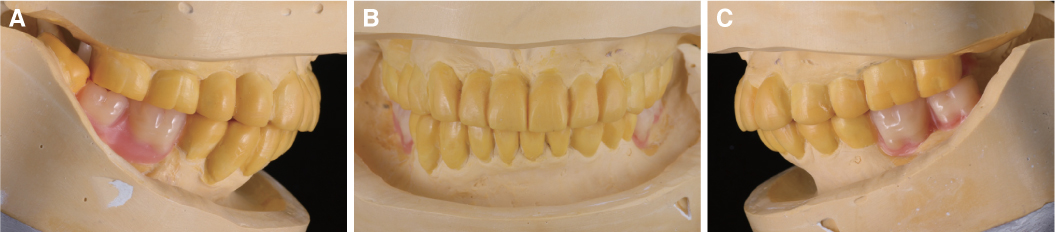
Fig. 4 Diagnostic wax-up. (A) Right lateral view, (B) Frontal view, (C) Left lateral view are shown.
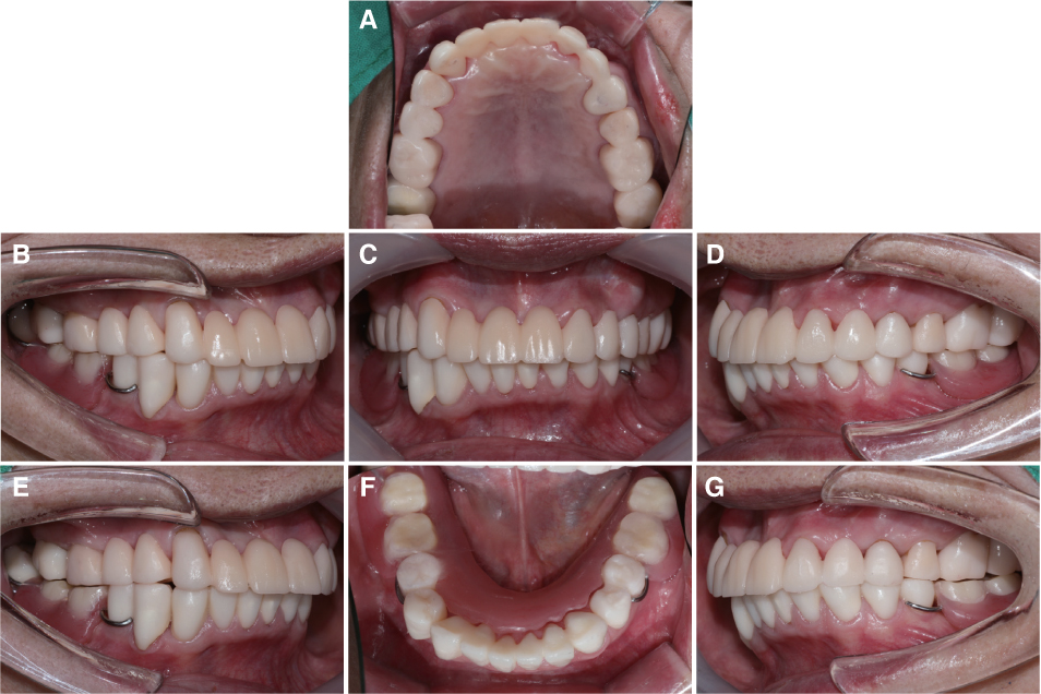
Fig. 5 Intraoral photographs and eccentric guidance of provisional restoration. (A) Maxillary occlusal view, (B) Right lateral view, (C) Frontal view, (D) Left lateral view, (E) Group function at right side, (F) Mandibular occlusal view, (G) Group function at left side are shown.
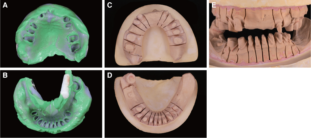
Fig. 6 Definitive impression taking and master cast. (A, B) Impression body, (C, D) Master cast, (E) Master cast mounted are shown.
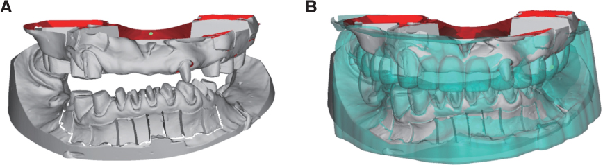
Fig. 7 Virtual master cast. (A) Superimposition of scanned separate dies and second die, (B) Superimposition of provisional cast and master cast using double scanning are shown.
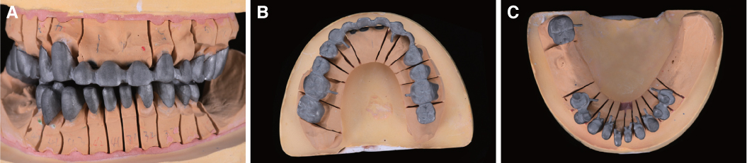
Fig. 8 Metal coping fabricated by 3D printing. (A) Frontal view, (B) Maxillary occlusal view, (C) Mandibular occlusal view are shown.
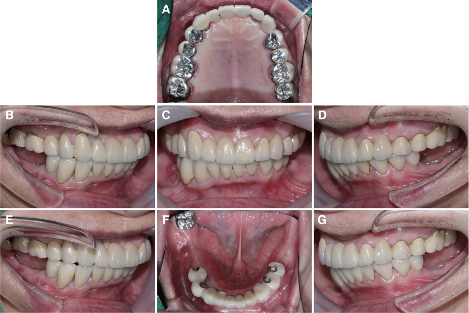
Fig. 9 Intraoral photographs and eccentric guidance of definitive fixed restoration. (A) Maxillary occlusal view, (B) Right lateral view, (C) Frontal view, (D) Left lateral view, (E) Group function at right side, (F) Mandibular occlusal view, (G) Group function at left side are shown.
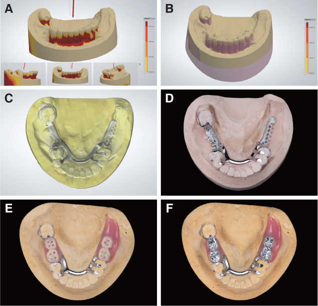
Fig. 10 Removable partial denture (RPD) fabrication procedures. (A) Electronic surveying of master cast, (B) Block out with respect to selected insertion path, (C) RPD framework was designed in CAD software, (D) Metal framework, (E) Occlusal reduction and retention hole formation, (F) RPD with metal occlusion are shown.
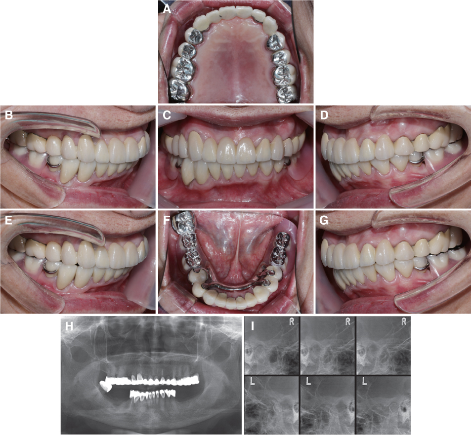
Fig. 11 Postoperative intraoral photographs and radiographic evaluation. (A) Maxillary occlusal view, (B) Right lateral view, (C) Frontal view, (D) Left lateral view, (E) Group function at right side, (F) Mandibular occlusal view, (G) Group function at left side, (H) Panoramic radiograph, (I) Transcranial view are shown.
Reference
-
1. Al Maaz A, Thompson GA, Drago C, An H, Berzins D. Effect of finish line design and metal alloy on the marginal and internal gaps of selective laser melting printed copings. J Prosthet Dent. 2019; 122:143–151.
Article2. Wang W, Yu H, Liu Y, Jiang X, Gao B. Trueness analysis of zirconia crowns fabricated with 3-dimensional printing. J Prosthet Dent. 2019; 121:285–291.
Article3. Arnold C, Hey J, Schweyen R, Setz JM. Accuracy of CAD/CAM-fabricated removable partial dentures. J Prosthet Dent. 2018; 119:586–592.
Article4. Padwa BL, Kaiser MO, Kaban LB. Occlusal cant in the frontal plane as a reflection of facial asymmetry. J Oral Maxillofac Surg. 1997; 55:811–816.
Article5. Pinho T, Neves M, Alves C. Multidisciplinary management including periodontics, orthodontics, implants, and prosthetics for an adult. Am J Orthod Dentofacial Orthop. 2012; 142:235–245.
Article6. Ramfjord SP, Blankenship JR. Increased occlusal vertical dimension in adult monkeys. J Prosthet Dent. 1981; 45:74–83.
Article7. Dawson PE. Functional occlusion: From TMJ to smile design. St Louis: Mosby Elsevier;2007.8. Hemmings KW, Darbar UR, Vaughan S. Tooth wear treated with direct composite restorations at an increased vertical dimension: results at 30 months. J Prosthet Dent. 2000; 83:287–293.
Article9. Sato S, Hotta TH, Pedrazzi V. Removable occlusal overlay splint in the management of tooth wear: a clinical report. J Prosthet Dent. 2000; 83:392–395.
Article10. Smith BG, Knight JK. A comparison of patterns of tooth wear with aetiological factors. Br Dent J. 1984; 157:16–19.
Article11. Shifman A, Laufer BZ, Chweidan H. Posterior bite collapse-revisited. J Oral Rehabil. 1998; 25:376–385.12. Turner KA, Missirlian DM. Restoration of the extremely worn dentition. J Prosthet Dent. 1984; 52:467–474.
Article13. Abduo J, Lyons K. Clinical considerations for increasing occlusal vertical dimension: a review. Aust Dent J. 2012; 57:2–10.
Article14. Karl M, Graef F, Wichmann M, Krafft T. Passivity of fit of CAD/CAM and copy-milled frameworks, veneered frameworks, and anatomically contoured, zirconia ceramic, implant-supported fixed prostheses. J Prosthet Dent. 2012; 107:232–238.
Article15. Kim SB, Kim NH, Kim JH, Moon HS. Evaluation of the fit of metal copings fabricated using stereolithography. J Prosthet Dent. 2018; 120:693–698.
Article16. Fathi HM, Al-Masoody AH, El-Ghezawi N, Johnson A. The accuracy of fit of crowns made from wax patterns produced conventionally (hand formed) and via CAD/CAM technology. Eur J Prosthodont Restor Dent. 2016; 24:10–17.17. Pjetursson BE, Tan K, Lang NP, Brägger U, Egger M, Zwahlen M. A systematic review of the survival and complication rates of fixed partial dentures (FPDs) after an observation period of at least 5 years. Clin Oral Implants Res. 2004; 15:667–676.
Article18. Dawson PE. Determining the determinants of occlusion. Int J Periodontics Restorative Dent. 1983; 3:8–21.19. Proffit WR. Equilibrium theory revisited: factors influencing position of the teeth. Angle Orthod. 1978; 48:175–186.20. Carlsson GE, Ingervall B, Kocak G. Effect of increasing vertical dimension on the masticatory system in subjects with natural teeth. J Prosthet Dent. 1979; 41:284–289.
Article
- Full Text Links
-
- Actions
-
Cited
- CITED
-
- Close
- Share
-
- Similar articles
-
- Full-mouth rehabilitation of a patient with loss of posterior support and collapsed occlusion utilizing dental CAD-CAM system
- Full mouth rehabilitation of a severely worn dentition using intraoral scanner and the CAD/CAM double scanning technique
- Full-mouth rehabilitation in an amelogenesis imperfecta patient with anterior open bite using CAD/CAM system
- Oral rehabilitation of a patient with severely worn dentition using monolithic zirconia
- Full mouth rehabilitation with vertical dimension increase in patient with severly worn dentition
