Intramuscular hematoma on the psoas muscle
- Affiliations
-
- 1Department of Neurosurgery, VHS Medical Center, Seoul, Korea. euro3399@naver.com
- KMID: 2461134
- DOI: http://doi.org/10.13004/kjnt.2019.15.e29
Abstract
- Intramuscular hematomas on the psoas muscle are rare and usually occur as a result of trauma, iatrogenic etiology during lumbar surgery, rupture of the aortic aneurysm, and hematologic diseases. The incidence of spontaneous psoas muscle hematomas has slowly increased as a result of using anticoagulation and antiplatelet agents. Magnetic resonance (MR) imaging is a more sensitive option compared to computed tomography (CT) when diagnosing a hematoma. Coronal T2-weighted images are more useful. CT imaging is also useful to establish the rapid diagnosis of hematoma. When a prolonged prothrombin time and international normalized ratio and decrease platelet count are noted, psoas muscle hematomas should be considered, if there was no lesion in the spinal canal. Most hematomas resolve spontaneously without clinical complications if the hematoma is not large or it is not compressing the surrounding important structures, irrespective of cause.
MeSH Terms
Figure
-
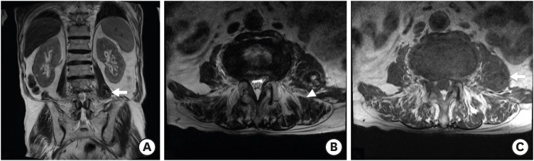
FIGURE 1 Magnetic resonance imaging reveals an oval-shaped mass lesion in the left psoas muscle. T2 coronal (A, white arrow), T2 axial (B), and T1 axial (C, white arrow) image. Various signal intensities within the hematoma, named mosaic sign (B, white arrow head).
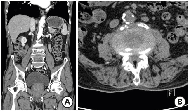
FIGURE 2 Three weeks later, computed tomography imaging reveals nearly complete absorption of the hematoma. Coronal (A) and axial (B) image.
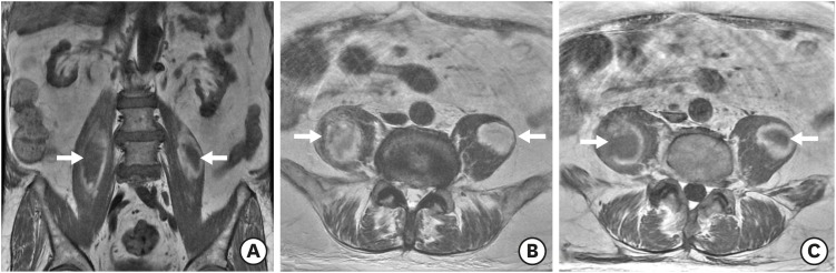
FIGURE 3 Magnetic resonance imaging without enhancement shows heterogeneous signal intensity mass with peripheral high intensity rim in both psoas muscles. T1 coronal (A, white arrows), T2 axial (B, white arrows), and T1 axial (C, white arrows) image.
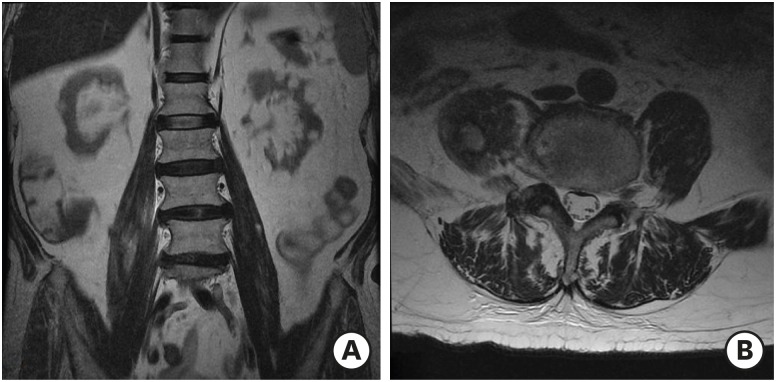
FIGURE 4 Four weeks later, magnetic resonance imaging reveals decreased hematoma size in both psoas muscles. T2 coronal (A) and T2 axial (B) image.
Reference
-
1. Akata S, Ohkubo Y, Jinho P, Saito K, Yamagishi T, Yoshimura M, et al. MR features of a case of chronic expanding hematoma. Clin Imaging. 2000; 24:44–46. PMID: 11120417.
Article2. Almushayti Z. Hematoma of the psoas muscle, in prostatic cancer patient: a case report. Pan Afr Med J. 2015; 20:138. PMID: 27386016.
Article3. Basheer A, Jain R, Anton T, Rock J. Bilateral iliopsoas hematoma: case report and literature review. Surg Neurol Int. 2013; 4:121. PMID: 24232386.
Article4. Cronin CG, Lohan DG, Meehan CP, Delappe E, McLoughlin R, O'Sullivan GJ, et al. Anatomy, pathology, imaging and intervention of the iliopsoas muscle revisited. Emerg Radiol. 2008; 15:295–310. PMID: 18548299.
Article5. Kim HS, Ju CI, Kim SW, Kim JG. Huge psoas muscle hematoma due to lumbar segmental vessel injury following percutaneous endoscopic lumbar discectomy. J Korean Neurosurg Soc. 2009; 45:192–195. PMID: 19352485.
Article6. Lakkol S, Sarda P, Karpe P, Krishna M. Conservative management of psoas haematoma following complex lumbar surgery. Indian J Orthop. 2014; 48:107–110. PMID: 24600073.
Article7. Lee JK, Glazer HS. Psoas muscle disorders: MR imaging. Radiology. 1986; 160:683–687. PMID: 3737906.
Article8. Lefevre N, Bohu Y, Klouche S, Chemla N, Herman S. Complete paralysis of the quadriceps secondary to post-traumatic iliopsoas hematoma: a systematic review. Eur J Orthop Surg Traumatol. 2015; 25:39–43. PMID: 23996110.
Article9. Lenchik L, Dovgan DJ, Kier R. CT of the iliopsoas compartment: value in differentiating tumor, abscess, and hematoma. AJR Am J Roentgenol. 1994; 162:83–86. PMID: 8273696.
Article10. Marquardt G, Barduzal Angles S, Leheta F, Seifert V. Spontaneous haematoma of the iliac psoas muscle: a case report and review of the literature. Arch Orthop Trauma Surg. 2002; 122:109–111. PMID: 11880914.11. Robinson DE, Ball KE, Webb PJ. Iliopsoas hematoma with femoral neuropathy presenting a diagnostic dilemma after spinal decompression. Spine. 2001; 26:E135–E138. PMID: 11246396.
Article12. Saad Z, Ahmed B, Mostafa R, Hicham B, Lahcen B. Conservative treatment of a psoas hematoma revealed by a lower limb palsy. Pan Afr Med J. 2017; 28:138. PMID: 29541288.
Article13. Sarwat AM, Sutcliffe JC. Haematoma of the psoas muscle after posterior spinal instrumentation and enoxaparin prophylaxis. Grand Rounds. 2003; 3:35–37.
- Full Text Links
-
- Actions
-
Cited
- CITED
-
- Close
- Share
-
- Similar articles
-
- Ultrasonographic Findings of Psoas abscess and Hematoma
- Psoas sign: a reevaluation
- Delayed psoas muscle hematoma formation after spinal anesthesia with the paramedian approach in a hemodialysis patient: A case report
- A Case of Occult Retroperitoneal Hematoma Associated with Head Trauma
- Non-Hodgkin Lymphoma Occurred in Psoas Muscle
