Cervical Cerebrospinal Fluid Leakage Concomitant with a Thoracic Spinal Intradural Arachnoid Cyst
- Affiliations
-
- 1Department of Neurosurgery, Chungnam National University Hospital, School of Medicine, Chungnam National University, Daejeon, Korea. swchoi@cnu.ac.kr
- KMID: 2461131
- DOI: http://doi.org/10.13004/kjnt.2019.15.e31
Abstract
- We encountered a very rare case of spontaneous spinal cerebrospinal fluid (CSF) leakage and a spinal intradural arachnoid cyst (AC) that were diagnosed at different sites in the same patient. These two lesions were thought to have interfered with the disease onset and deterioration. A 30-year-old man presented with sudden neck pain and orthostatic headache. Diplopia, ophthalmic pain, and headache deteriorated. CSF leakage was confirmed in C2 by radioisotope cisternography, and an epidural blood patch was performed. While his symptoms improved gradually, paraparesis suddenly progressed. Thoracolumbar magnetic resonance imaging (MRI) revealed an upper thoracic spinal intradural AC, which was compressing the spinal cord. We removed the outer membrane of the AC and performed fenestration of the inner membrane after T3-4 laminectomy. Postoperative MRI showed complete removal of the AC and normalized lumbar subarachnoid space. All neurological deficits including motor weakness, sensory impairment, and voiding function improved to normal. We present a case of spontaneous spinal CSF leakage and consecutive intracranial hypotension in a patient with a spinal AC. Our report suggests that if spinal CSF leakage and a spinal AC are diagnosed in one patient, even if they are located at different sites, they may affect disease progression and aggravation.
MeSH Terms
Figure
-
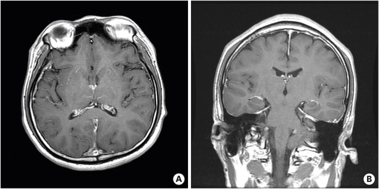
FIGURE 1 Magnetic resonance images showed mild contrast enhancement of meninges.
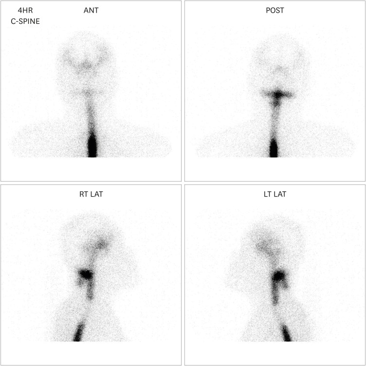
FIGURE 2 Radioisotope cisternography revealed cerebrospinal fluid leakage at the C2 level.ANT: anterior, POST: posterior, RT LAT: right lateral, LT LAT: left lateral.
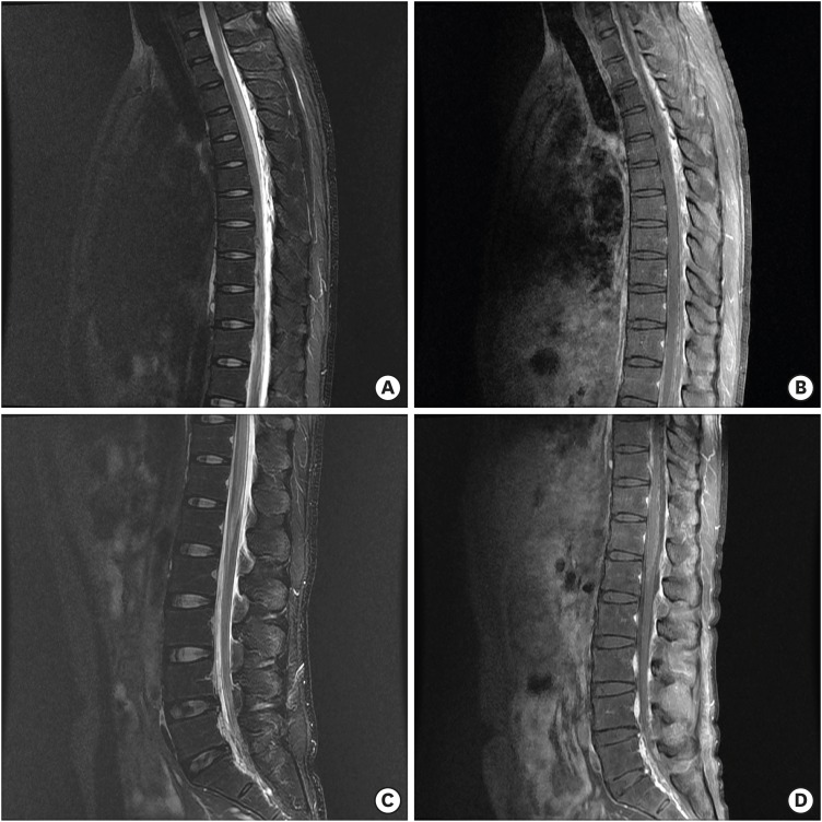
FIGURE 3 Thoracolumbar magnetic resonance imaging showed mild expansion of the subarachnoid space at the upper thoracic levels with contrast enhancement of the arachnoid membrane (A, B). In contrast, the lumbar subarachnoid space became narrow (C, D).
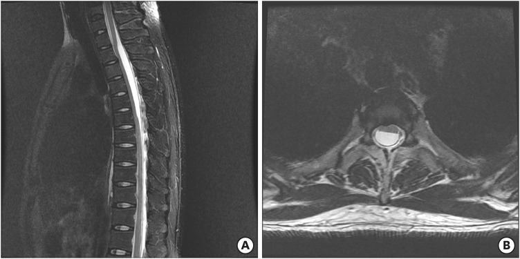
FIGURE 4 Magnetic resonance imaging revealed an intradural arachnoid cyst severely compressing the spinal cord at the upper thoracic spine.
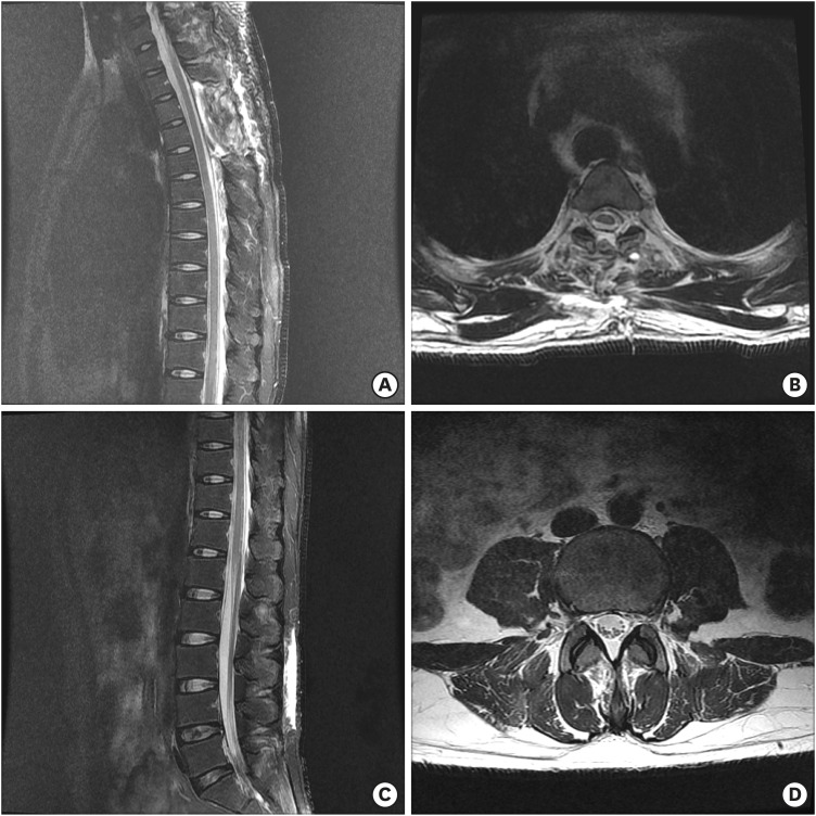
FIGURE 5 Postoperative magnetic resonance imaging showed that arachnoid cyst was removed completely, compression of the spinal cord disappeared, and the subarachnoid space at the lumbar levels was normalized.
Reference
-
1. Bond AE, Zada G, Bowen I, McComb JG, Krieger MD. Spinal arachnoid cysts in the pediatric population: report of 31 cases and a review of the literature. J Neurosurg Pediatr. 2012; 9:432–441. PMID: 22462711.
Article2. Chan SM, Chodakiewitz YG, Maya MM, Schievink WI, Moser FG. Intracranial hypotension and cerebrospinal fluid leak. Neuroimaging Clin N Am. 2019; 29:213–226. PMID: 30926112.
Article3. Ergun E, Börcek AO, Cemil B, Doğulu F, Baykaner MK. Should we operate all extradural spinal arachnoid cysts? Report of a case. Turk Neurosurg. 2008; 18:52–55. PMID: 18382979.4. Eroglu U, Bozkurt M, Kahilogullari G, Dogan I, Ozgural O, Shah KJ, et al. Surgical management of spinal arachnoid cysts in adults. World Neurosurg. 2019; 122:e1146–e1152. PMID: 30447456.
Article5. Ferrante E, Trimboli M, Rubino F. Spontaneous intracranial hypotension: review and expert opinion. Acta Neurol Belg. 2019.
Article6. Lin JP, Zhang SD, He FF, Liu MJ, Ma XX. The status of diagnosis and treatment to intracranial hypotension, including SIH. J Headache Pain. 2017; 18:4. PMID: 28091819.
Article7. Mokri B, Maher CO, Sencakova D. Spontaneous CSF leaks: underlying disorder of connective tissue. Neurology. 2002; 58:814–816. PMID: 11889250.
Article8. Rincon F, Mocco J, Komotar RJ, Khandji AG, McCormick PC, Olarte M. Chronic myelopathy due to a giant spinal arachnoid cyst: a complication of the intrathecal injection of phenol. J Neurosurg Spine. 2008; 8:390–393. PMID: 18377326.
Article9. Sadek AR, Nader-Sepahi A. Spinal arachnoid cysts: presentation, management and pathophysiology. Clin Neurol Neurosurg. 2019; 180:87–96. PMID: 30952036.
Article10. Schievink WI. Spontaneous spinal cerebrospinal fluid leaks and intracranial hypotension. JAMA. 2006; 295:2286–2296. PMID: 16705110.
Article11. Siedler DG, Ibbett IM, Thani NB. Surgical management of spontaneous spinal cerebrospinal fluid epidural fistula. World Neurosurg. 2017; 99:810.e5–810.e10.
Article12. Upadhyaya P, Ailani J. A review of spontaneous intracranial hypotension. Curr Neurol Neurosci Rep. 2019; 19:22. PMID: 30888542.
Article13. Wang MY, Levi AD, Green BA. Intradural spinal arachnoid cysts in adults. Surg Neurol. 2003; 60:49–55. PMID: 12865013.
Article14. Kim YS, Cho BM, Cho SM, Park SH, Oh SM. Two cases of secondary intracranial hypotension. J Korean Neurotraumatol Soc. 2011; 7:103–107.
Article
