Korean J Neurotrauma.
2019 Oct;15(2):187-191. 10.13004/kjnt.2019.15.e22.
Presyrinx Associated with Post-Traumatic Hydrocephalus Successfully Treated by Ventriculoperitoneal Shunt
- Affiliations
-
- 1Department of Neurosurgery, Soonchunhyang University Bucheon Hospital, Bucheon, Korea. sunchulh@schmc.ac.kr
- KMID: 2461126
- DOI: http://doi.org/10.13004/kjnt.2019.15.e22
Abstract
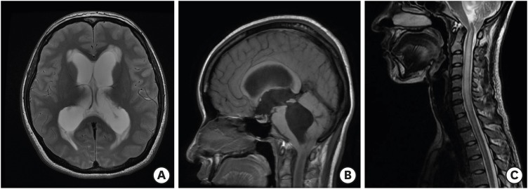
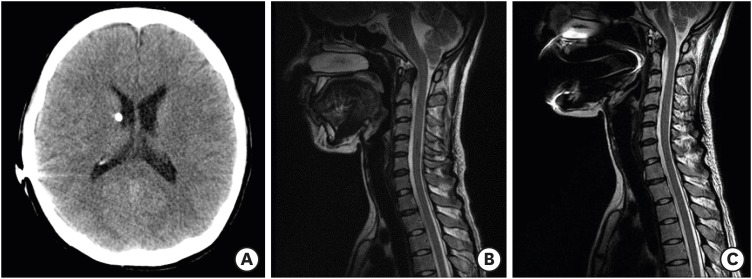
- Presyrinx consists of reversible spinal cord swelling without frank cavitation, as observed on T2 weighted magnetic resonance imaging (MRI). The condition may evolve into syringomyelia, but timely surgical interventions have achieved meaningful results. Here, we report the case of a 27-year-old woman who presented with headache, dizziness, and diplopia 2 months after suffering a mild head trauma. On MRI, hydrocephalus, downward herniation of the cerebellar tonsil, and a diffuse high signal change in the cervical spinal cord were detected. After insertion of a ventriculoperitoneal shunt, her neurological symptoms resolved, and she has had no signs of presyrinx recurrence for >4 years.
MeSH Terms
Figure
Reference
-
1. Cardoso ER, Galbraith S. Posttraumatic hydrocephalus--a retrospective review. Surg Neurol. 1985; 23:261–264. PMID: 3975808.
Article2. Daou B, Klinge P, Tjoumakaris S, Rosenwasser RH, Jabbour P. Revisiting secondary normal pressure hydrocephalus: does it exist? A review. Neurosurg Focus. 2016; 41:E6.
Article3. Fischbein NJ, Dillon WP, Cobbs C, Weinstein PR. The “presyrinx” state: a reversible myelopathic condition that may precede syringomyelia. AJNR Am J Neuroradiol. 1999; 20:7–20. PMID: 9974051.4. Gardner WJ, Angel J. The mechanism of syringomyelia and its surgical correction. Clin Neurosurg. 1958; 6:131–140. PMID: 13826542.
Article5. Goh S, Bottrell CL, Aiken AH, Dillon WP, Wu YW. Presyrinx in children with Chiari malformations. Neurology. 2008; 71:351–356. PMID: 18565831.
Article6. Greitz D. Unraveling the riddle of syringomyelia. Neurosurg Rev. 2006; 29:251–263. PMID: 16752160.
Article7. Hakim S, Adams RD. The special clinical problem of symptomatic hydrocephalus with normal cerebrospinal fluid pressure. Observations on cerebrospinal fluid hydrodynamics. J Neurol Sci. 1965; 2:307–327. PMID: 5889177.8. Levy EI, Heiss JD, Kent MS, Riedel CJ, Oldfield EH. Spinal cord swelling preceding syrinx development. Case report. J Neurosurg. 2000; 92:93–97. PMID: 10616064.9. Milhorat TH, Kotzen RM. Stenosis of the central canal of the spinal cord following inoculation of suckling hamsters with reovirus type I. J Neurosurg. 1994; 81:103–106. PMID: 8207510.
Article10. Milhorat TH, Capocelli AL Jr, Anzil AP, Kotzen RM, Milhorat RH. Pathological basis of spinal cord cavitation in syringomyelia: analysis of 105 autopsy cases. J Neurosurg. 1995; 82:802–812. PMID: 7714606.
Article11. Muthukumar N, Venkatesh G, Thiruppathy S. Arrested hydrocephalus and the presyrinx state. Case report. J Neurosurg. 2005; 103:466–470. PMID: 16302623.12. Muthukumar N. Preysyrinx state and shunt dysfunction: an underrecognized entity? Acta Neurochir (Wien). 2010; 152:1969–1973. PMID: 20669036.13. Nomura S, Akimura T, Imoto H, Nishizaki T, Suzuki M. Endoscopic fenestration of posterior fossa arachnoid cyst for the treatment of presyrinx myelopathy--case report. Neurol Med Chir (Tokyo). 2002; 42:452–454. PMID: 12416571.14. Oldfield EH, Muraszko K, Shawker TH, Patronas NJ. Pathophysiology of syringomyelia associated with Chiari I malformation of the cerebellar tonsils. Implications for diagnosis and treatment. J Neurosurg. 1994; 80:3–15. PMID: 8271018.15. Takaishi Y, Suzuki T, Iwakura M, Mizukawa K, Yasuo K, Matsumoto S. Presyrinx state with Chiari malformation. Spinal Surg. 2009; 23:77–79.16. Wan MJ, Nomura H, Tator CH. Conversion to symptomatic Chiari I malformation after minor head or neck trauma. Neurosurgery. 2008; 63:748–753. PMID: 18981886.
Article17. Wang R, Qiu Y, Jiang J, Liu Z. Atypical syringomyelia without cavity in a patient with Chiari malformation and hydrocephalus. Spine (Phila Pa 1976). 2007; 32:E467–E470. PMID: 17632386.
Article
- Full Text Links
- Actions
-
Cited
- CITED
-
- Close
- Share
- Similar articles
-
- Asymptomatic Fracture of a Distal Shunt Catheter after Ventriculoperitoneal Shunt for Post-traumatic Hydrocephalus
- Isolated Lateral Ventricle after V-P Shunt
- Ventriculoperitoneal Shunt Malfunction during Pregnancy
- Perforation of the Urinary Bladder by the Distal Catheter: A Rare Complication of Ventriculoperitoneal Shunt
- A Case of Post-Traumatic Pseudomeningocele Treated by Lumboperitoneal Shunt


