Surface alterations following instrumentation with a nylon or metal brush evaluated with confocal microscopy
- Affiliations
-
- 1Department of Dentistry, University of Ulsan College of Medicine, Seoul, Korea.
- 2Department of Periodontics, Asan Medical Center, Seoul, Korea.
- 3Department of Periodontics, Seoul St. Mary's Hospital, The Catholic University of Korea College of Medicine, Seoul, Korea. ko_y@catholic.ac.kr
- KMID: 2461021
- DOI: http://doi.org/10.5051/jpis.2019.49.5.310
Abstract
- PURPOSE
Surface alterations of titanium discs following instrumentation with either a nylon brush or a metal brush were evaluated.
METHODS
A total of 27 titanium discs with 3 surface types (9 discs for each type), including machined (M) surfaces, sandblasted and acid-etched (SA) surfaces, and surfaces treated by resorbable blast media (RBM), were used. Three discs were instrumented with a nylon brush, another 3 discs were instrumented with a metal brush, and the remaining 3 discs were used as controls for each surface type. Surface properties including the arithmetic mean value of a linear profile (Ra), maximum height of a linear profile (Rz), skewness of the assessed linear profile (Rsk), arithmetic mean height of a surface (Sa), maximum height of a surface (Sz), developed interfacial area ratio (Sdr), skewness of a surface profile (Ssk), and kurtosis of a surface profile (Sku) were measured using confocal microscopy.
RESULTS
Instrumentation with the nylon brush increased the Ra, Sa, and Sdr of the M surfaces. On the SA surfaces, Ra, Sa and Sdr decreased after nylon brush use. Meanwhile, the roughness of the RBM surface was not affected by the nylon brush. The use of the metal brush also increased the Ra, Sa, and Sdr of the M surface; however, the increase in Sdr was not statistically significant (P=0.119). The decreases in the Rz, Sz, Ra, Sa, and Sdr of the SA surfaces were remarkable. On the RBM surfaces, the use of the metal brush did not cause changes in Ra and Sa, whereas Rz, Sz, and Sdr were reduced.
CONCLUSIONS
Titanium surfaces were altered when instrumented either with a nylon brush or a metal brush. Hence, it is recommended that nylon or metal brushes be used with caution in order to avoid damaging the implant fixture/abutment surface.
Keyword
MeSH Terms
Figure
-

Figure 1 The instruments used in the present study. The nylon brush (left) had a shape similar to a tapered interdental brush and it should be used with an equipment (not shown in this picture) provided by the manufacturer. The metal brush (right) is a low-speed rotary bur with laterally protruding stainless steel bristles at its end.
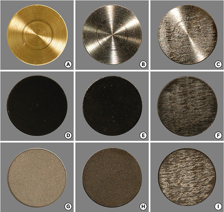
Figure 2 Gross view of titanium discs. (A) M surface without instrumentation, (B) M surface instrumented with a nylon brush, (C) M surface instrumented with a metal brush, (D) SA surface without instrumentation, (E) SA surface instrumented with a nylon brush, (F) SA surface instrumented with a metal brush, (G) surface treated with RBM without instrumentation, (H) RBM surface instrumented with a nylon brush, (I) RBM surface instrumented with a metal brush. The changes on the surfaces instrumented with a nylon brush were indistinct (B, E, and H) and the alterations on the surfaces instrumented with a metal brush were obvious (C, F, and I). M: machined, SA: sandblasted and acid-etched, RBM: resorbable blast media.

Figure 3 Images from intensity projections with an extended depth of focus obtained by confocal laser microscopy. (A) M surface without instrumentation, (B) M surface instrumented with a nylon brush, (C) M surface instrumented with a metal brush, (D) SA surface without instrumentation, (E) SA surface instrumented with a nylon brush, (F) SA surface instrumented with a metal brush, (G) surface treated with RBM without instrumentation, (H) RBM surface instrumented with a nylon brush, (I) RBM surface instrumented with a metal brush. Instrumentation with the metal brush resulted in scratches on the M surface (C) and flattening of rough surfaces (F and I). M: machined, SA: sandblasted and acid-etched, RBM: resorbable blast media.
Reference
-
1. Zitzmann NU, Berglundh T. Definition and prevalence of peri-implant diseases. J Clin Periodontol. 2008; 35:286–291.
Article2. Mombelli A, Lang NP. The diagnosis and treatment of peri-implantitis. Periodontol 2000. 1998; 17:63–76.
Article3. Quirynen M, van der Mei HC, Bollen CM, Schotte A, Marechal M, Doornbusch GI, et al. An in vivo study of the influence of the surface roughness of implants on the microbiology of supra- and subgingival plaque. J Dent Res. 1993; 72:1304–1309.
Article4. Rimondini L, Farè S, Brambilla E, Felloni A, Consonni C, Brossa F, et al. The effect of surface roughness on early in vivo plaque colonization on titanium. J Periodontol. 1997; 68:556–562.
Article5. Bollen CM, Lambrechts P, Quirynen M. Comparison of surface roughness of oral hard materials to the threshold surface roughness for bacterial plaque retention: a review of the literature. Dent Mater. 1997; 13:258–269.
Article6. Bollen CM, Papaioanno W, Van Eldere J, Schepers E, Quirynen M, van Steenberghe D. The influence of abutment surface roughness on plaque accumulation and peri-implant mucositis. Clin Oral Implants Res. 1996; 7:201–211.
Article7. Quirynen M, Bollen CM, Papaioannou W, Van Eldere J, van Steenberghe D. The influence of titanium abutment surface roughness on plaque accumulation and gingivitis: short-term observations. Int J Oral Maxillofac Implants. 1996; 11:169–178.8. Alhag M, Renvert S, Polyzois I, Claffey N. Re-osseointegration on rough implant surfaces previously coated with bacterial biofilm: an experimental study in the dog. Clin Oral Implants Res. 2008; 19:182–187.
Article9. Park JB, Jang YJ, Choi BK, Kim KK, Ko Y. Treatment with various ultrasonic scaler tips affects efficiency of brushing of SLA titanium discs. J Craniofac Surg. 2013; 24:e119–23.
Article10. Park JB, Jang YJ, Koh M, Choi BK, Kim KK, Ko Y. In vitro analysis of the efficacy of ultrasonic scalers and a toothbrush for removing bacteria from resorbable blast material titanium disks. J Periodontol. 2013; 84:1191–1198.
Article11. Park JB, Kim N, Ko Y. Effects of ultrasonic scaler tips and toothbrush on titanium disc surfaces evaluated with confocal microscopy. J Craniofac Surg. 2012; 23:1552–1558.
Article12. Wohlfahrt JC, Lyngstadaas SP. Mechanical debridement of a peri-implant osseous defect with a novel titanium brush and reconstruction with porous titanium granules: a case report with reentry surgery. Clin Adv Periodontics. 2012; 2:136–140.
Article13. Schmidt KE, Auschill TM, Heumann C, Frankenberger R, Eick S, Sculean A, et al. Clinical and laboratory evaluation of the effects of different treatment modalities on titanium healing caps: a randomized, controlled clinical trial. Clin Oral Investig. 2018; 22:2149–2160.
Article14. Bertoldi C, Lusuardi D, Battarra F, Sassatelli P, Spinato S, Zaffe D. The maintenance of inserted titanium implants: in-vitro evaluation of exposed surfaces cleaned with three different instruments. Clin Oral Implants Res. 2017; 28:57–63.
Article15. Chen CJ, Ding SJ, Chen CC. Effects of surface conditions of titanium dental implants on bacterial adhesion. Photomed Laser Surg. 2016; 34:379–388.
Article16. Sahrmann P, Ronay V, Hofer D, Attin T, Jung RE, Schmidlin PR. In vitro cleaning potential of three different implant debridement methods. Clin Oral Implants Res. 2015; 26:314–319.
Article17. Fox SC, Moriarty JD, Kusy RP. The effects of scaling a titanium implant surface with metal and plastic instruments: an in vitro study. J Periodontol. 1990; 61:485–490.
Article18. Homiak AW, Cook PA, DeBoer J. Effect of hygiene instrumentation on titanium abutments: a scanning electron microscopy study. J Prosthet Dent. 1992; 67:364–369.
Article19. Rühling A, Kocher T, Kreusch J, Plagmann HC. Treatment of subgingival implant surfaces with Teflon-coated sonic and ultrasonic scaler tips and various implant curettes. An in vitro study. Clin Oral Implants Res. 1994; 5:19–29.
Article20. Meschenmoser A, d’Hoedt B, Meyle J, Elssner G, Korn D, Hämmerle H, et al. Effects of various hygiene procedures on the surface characteristics of titanium abutments. J Periodontol. 1996; 67:229–235.
Article21. Schmage P, Thielemann J, Nergiz I, Scorziello TM, Pfeiffer P. Effects of 10 cleaning instruments on four different implant surfaces. Int J Oral Maxillofac Implants. 2012; 27:308–317.22. Mengel R, Meer C, Flores-de-Jacoby L. The treatment of uncoated and titanium nitride-coated abutments with different instruments. Int J Oral Maxillofac Implants. 2004; 19:232–238.23. Louropoulou A, Slot DE, Van der Weijden FA. Titanium surface alterations following the use of different mechanical instruments: a systematic review. Clin Oral Implants Res. 2012; 23:643–658.
Article24. Evans AR, Harper IS, Sanson GD. Confocal imaging, visualization and 3-D surface measurement of small mammalian teeth. J Microsc. 2001; 204:108–118.
Article25. Howell K, Hopkins N, Mcloughlin P. Combined confocal microscopy and stereology: a highly efficient and unbiased approach to quantitative structural measurement in tissues. Exp Physiol. 2002; 87:747–756.
Article26. Kwan JY, Zablotsky MH, Meffert RM. Implant maintenance using a modified ultrasonic instrument. J Dent Hyg. 1990; 64:422.27. Rapley JW, Swan RH, Hallmon WW, Mills MP. The surface characteristics produced by various oral hygiene instruments and materials on titanium implant abutments. Int J Oral Maxillofac Implants. 1990; 5:47–52.28. Rimondini L, Cicognani Simoncini F, Carrassi A. Micro-morphometric assessment of titanium plasma-sprayed coating removal using burs for the treatment of peri-implant disease. Clin Oral Implants Res. 2000; 11:129–138.
Article29. Rühling A, Hellweg A, Plagmann HC, Kocher T. Removal of HA and TPS implant coatings and fibroblast attachment on exposed surfaces. Clin Oral Implants Res. 2001; 12:301–308.
Article30. Barbour ME, O'Sullivan DJ, Jenkinson HF, Jagger DC. The effects of polishing methods on surface morphology, roughness and bacterial colonisation of titanium abutments. J Mater Sci Mater Med. 2007; 18:1439–1447.
Article31. Duarte PM, Reis AF, de Freitas PM, Ota-Tsuzuki C. Bacterial adhesion on smooth and rough titanium surfaces after treatment with different instruments. J Periodontol. 2009; 80:1824–1832.
Article32. Schwarz F, Papanicolau P, Rothamel D, Beck B, Herten M, Becker J. Influence of plaque biofilm removal on reestablishment of the biocompatibility of contaminated titanium surfaces. J Biomed Mater Res A. 2006; 77A:437–444.
Article33. Bailey GM, Gardner JS, Day MH, Kovanda BJ. Implant surface alterations from a nonmetallic ultrasonic tip. J West Soc Periodontol Periodontal Abstr. 1998; 46:69–73.
- Full Text Links
-
- Actions
-
Cited
- CITED
-
- Close
- Share
-
- Similar articles
-
- Implant Surface Changes Observed with Confocal and Electron Microscopy After Probing with Metal and Plastic Periodontal Probes
- Inflammatory Vitiligo Confirmed Diagnosis and Treatment Outcome Using a Reflectance Confocal Microscopy
- Comparision of Specular Microscopy and Confocal Microscopy for Evaluation of Corneal Endothelium
- The Effects of a Er:YAG Laser on Machined, Sand-Blasted and Acid-Etched, and Resorbable Blast Media Titanium Surfaces Using Confocal Microscopy and Scanning Electron Microscopy
- In vivo tandem scanning confocal microscopy in acanthamoeba keratitis
