Investig Magn Reson Imaging.
2019 Sep;23(3):276-278. 10.13104/imri.2019.23.3.276.
Left-Sided Cisterna Chyli: a Case Report on a Rare Normal Anatomic Structure
- Affiliations
-
- 1Department of Radiology, Soonchunhyang University College of Medicine, Cheonan Hospital, Cheonan, Korea. jeongah.h09@gmail.com
- KMID: 2459883
- DOI: http://doi.org/10.13104/imri.2019.23.3.276
Abstract
- The cisterna chyli, a dilated lymphatic sac in the retrocrural space, is usually located to the right of the aorta. We report a case of a left-sided cisterna chyli, which was incidentally detected on the radiologic examinations of a preoperative workup for cholangiocarcinoma. Computed tomography (CT) and magnetic resonance (MR) images revealed a cisterna chyli measuring 2.5 cm in length in the left retrocrural space. The dilated lumbar lymphatics joined with the cisterna chyli, which was continuous with the left-sided thoracic duct. To the best of our knowledge, this is the second antemortem case of a left-sided cisterna chyli in literature. The cisterna chyli can mimic retrocrural lymphadenopathy, solid tumor with cystic degeneration, abscess or hematoma. The left-sided cisterna chyli should be referred to as a structure so as to be cautious in surgical approach.
MeSH Terms
Figure
-

Fig. 1 An incidentally-detected left-sided cisterna chyli in a 76-year-old man. Contrast enhanced CT image showed a well-defined non-enhancing lesion (arrow) measuring 1.2 × 1.4 × 2.5 cm of near-water attenuation. It was located in the left retrocrural space between the T12 and L1 vertebral level.
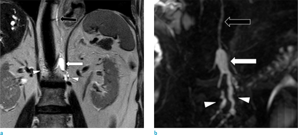
Fig. 2 MR coronal T2-weighted image (a) and coronal maximum intensity projection MR Cholangiopancreatography image (b) revealed dilated lumber lymphatics (arrowheads) converged to form a localized cystic structure demonstrating the classical appearance of a cisterna chyli (arrow). The cisterna chyli was situated in the left retrocrural space and was continuous with the left-sided thoracic duct (open arrows) at cephalad.
Reference
-
1. Rosenberger A, Abrams HL. Radiology of the thoracic duct. Am J Roentgenol Radium Ther Nucl Med. 1971; 111:807–820.
Article2. Meguid RA. Chylothorax: surgical ligation of the thoracic duct through thoracotomy. Oper Tech Thorac Cardiovasc Surg. 2016; 21:139–151.
Article3. Kurosaki Y, Fujikawa A. Left-sided cisterna chyli. AJR Am J Roentgenol. 2000; 175:1462.
Article4. Kiyonaga M, Mori H, Matsumoto S, Yamada Y, Sai M, Okada F. Thoracic duct and cisterna chyli: evaluation with multidetector row CT. Br J Radiol. 2012; 85:1052–1058.
Article5. Pinto PS, Sirlin CB, Andrade-Barreto OA, Brown MA, Mindelzun RE, Mattrey RF. Cisterna chyli at routine abdominal MR imaging: a normal anatomic structure in the retrocrural space. Radiographics. 2004; 24:809–817.
Article6. Lee KC, Cassar-Pullicino VN. Giant cisterna chyli: MRI depiction with gadolinium-DTPA enhancement. Clin Radiol. 2000; 55:51–55.
Article7. Gollub MJ, Castellino RA. The cisterna chyli: a potential mimic of retrocrural lymphadenopathy on CT scans. Radiology. 1996; 199:477–480.
Article8. Rha SE, Byun JY, Jung SE, Chun HJ, Lee HG, Lee JM. Neurogenic tumors in the abdomen: tumor types and imaging characteristics. Radiographics. 2003; 23:29–43.
Article9. Restrepo CS, Eraso A, Ocazionez D, Lemos J, Martinez S, Lemos DF. The diaphragmatic crura and retrocrural space: normal imaging appearance, variants, and pathologic conditions. Radiographics. 2008; 28:1289–1305.
Article
- Full Text Links
-
- Actions
-
Cited
- CITED
-
- Close
- Share
-
- Similar articles
-
- The Cisterna Chyli in Gastrointestinal Malignancy Patients: Incidence and Finding in CT
- Abdominal Organ Injuries with Chyloperitoneum after Blunt Tauma: A Case Report
- Laparoscopic cholecystectomy and common bile duct exploration for gallstone and common bile duct stone in a patient with a left-sided gallbladder: a case report
- Left-sided Gallbladder: 2 cases
- Left Sided Appendicitis in Patient with Intestinal Malrotation
