Cavernous Hemangioma of the Gallbladder: a Case Report
- Affiliations
-
- 1Department of Radiology, Jeju National University Hospital, Jeju National University School of Medicine, Jeju, Korea. shinshlee@naver.com
- 2Department of Pathology, Jeju National University Hospital, Jeju National University School of Medicine, Jeju, Korea.
- 3Department of Surgery, Jeju National University Hospital, Jeju National University School of Medicine, Jeju, Korea.
- KMID: 2459881
- DOI: http://doi.org/10.13104/imri.2019.23.3.264
Abstract
- Cavernous hemangioma of the gallbladder is an extremely rare benign tumor. The tumor has only a few cases being reported in literature. However, to the best of our knowledge, no reports focusing on the MRI findings of cavernous hemangioma of the gallbladder have been published. This study reports a case of gallbladder hemangioma with pathologic and radiologic reviews, including MRI findings.
Figure
-
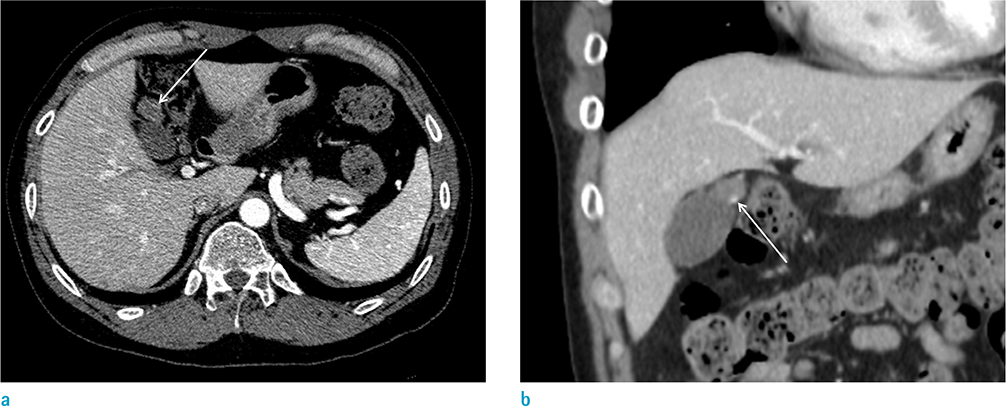
Fig. 1 A cavernous hemangioma of the gallbladder in a 53-year-old male patient, CT findings. (a, b) The axial (a) and coronal (b) contrast-enhanced CT images show a 1.8 cm, well-defined oval nodular lesion with a suspicious peripheral nodular enhancement (arrows) abutting the body of the gallbladder.
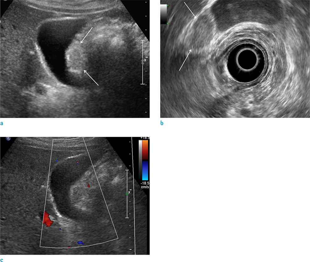
Fig. 2 A cavernous hemangioma of the gallbladder in a 53-year-old male patient, ultrasonographic findings. (a, b) Transabdominal ultrasonography (a) and endoscopic ultrasonography (b) demonstrated an oval shaped, isoechoic nodular lesion (arrows) with slightly hyperechoic rim. (c) Color Doppler examination did not reveal blood flow in the mass.
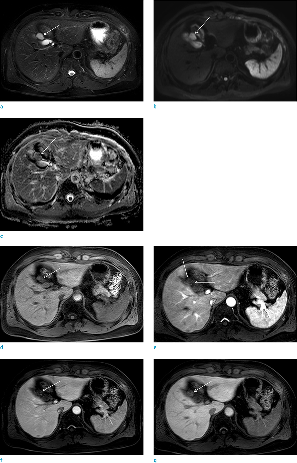
Fig. 3 A cavernous hemangioma of the gallbladder in a 53-year-old male patient, MRI findings. (a) The axial T2-weighted MR image demonstrates a well-defined nodular lesion with a homogeneous high signal intensity. (b) Diffusion weighted image with a high b value of 500 shows a high signal intensity lesion. (c) The corresponding lesion has slightly low value on apparent diffusion coefficient map. (d–g) The axial pre-contrast T1-weighted MR image (d), arterial phase (e), portal phase (f), and 3-minute delayed phase (g) show a small nodular lesion (arrows) with peripheral nodular and centripetal enhancement.
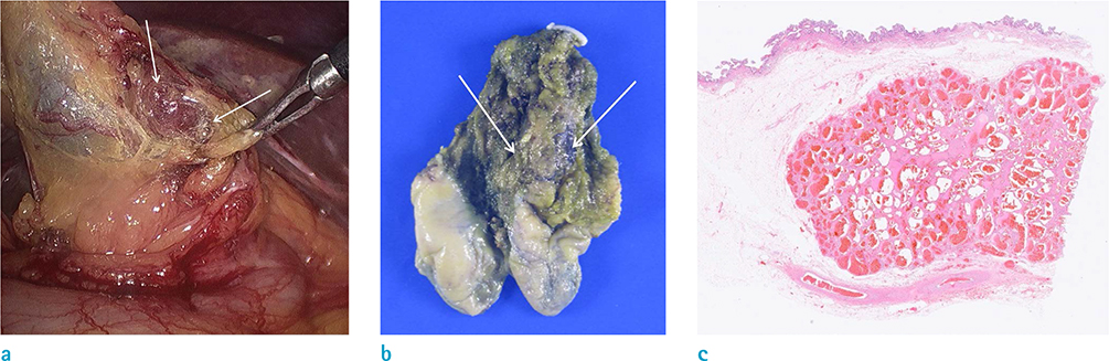
Fig. 4 A cavernous hemangioma of the gallbladder in a 53-year-old male patient. (a, b) In intraoperative finding (a) and gross examination (b), a well-demarcated dark reddish tumor (arrows) is identified on the wall of the gallbladder, originating from the sub-serosal perimuscular connective tissue. The tumor is completely enveloped by connective tissues. (c) Histopathology confirms the lesion as a cavernous hemangioma, which forms cystically dilated blood vessels lined by flat endothelial cells. There is no atypia (Hematoxylin & Eosin stain, × 5).
Reference
-
1. Crucitti A, La Greca A, Antinori A, Antonacci V, Magistrelli P. Cavernous hemangioma of the gallbladder. Case report and review of the literature. Tumori. 2005; 91:432–443.
Article2. Akama Y, Mizuguchi Y, Mamada Y, et al. A case of cavernous hemangioma of the gallbladder treated with single-incision laparoscopic cholecystectomy. Int Surg. 2016; 101:431–436.
Article3. Anderson SW, Kruskal JB, Kane RA. Benign hepatic tumors and iatrogenic pseudotumors. Radiographics. 2009; 29:211–229.
Article4. Melson GL, Reiter F, Evens RG. Tumorous conditions of the gallbladder. Semin Roentgenol. 1976; 11:269–282.
Article5. Jones WP, Keller FS, Odrezin GT, Kelly DR. Venous hemangioma of the gallbladder. Gastrointest Radiol. 1987; 12:319–321.
Article6. Catalano OA, Sahani DV, Kalva SP, et al. MR imaging of the gallbladder: a pictorial essay. Radiographics. 2008; 28:135–155. quiz 324.
Article7. Levy AD, Murakata LA, Rohrmann CA Jr. Gallbladder carcinoma: radiologic-pathologic correlation. Radiographics. 2001; 21:295–314. questionnaire, 549–255.
Article8. Choi WS, Kim SH, Lee ES, et al. CT findings of gallbladder metastases: emphasis on differences according to primary tumors. Korean J Radiol. 2014; 15:334–345.
Article9. Ishida M, Shiomi H, Naka S, Tani T, Okabe H. Leiomyoma of the gallbladder in a patient with metastatic gastrointestinal stromal tumor in the liver: a case report with differential diagnostic considerations. Oncol Lett. 2012; 4:1171–1173.
Article10. Botsford A, McKay K, Hartery A, Hapgood C. MRCP imaging of duplicate gallbladder: a case report and review of the literature. Surg Radiol Anat. 2015; 37:425–429.
Article
