Nervus terminalis and nerves to the vomeronasal organ: a study using human fetal specimens
- Affiliations
-
- 1Department of Anatomy, Wuxi School of Medicine, Jiangnan University, Wuxi, China.
- 2Department of Neurology, Wonkwang University School of Medicine and Hospital, Institute of Wonkwang Medical Science, Iksan, Korea. neurlogy@wonkwang.ac.kr, neuro20015@gmail.com
- 3Department of Maxillofacial Anatomy, Graduate School of Tokyo Medical and Dental University, Tokyo, Japan.
- 4Department of Anatomy, Tokyo Dental College, Tokyo, Japan.
- 5Sapporo Asuka Hospital, Sapporo, Japan.
- 6Department of Anatomy and Human Embryology, Institute of Embryology, Complutense University, Madrid, Spain.
- KMID: 2459543
- DOI: http://doi.org/10.5115/acb.19.020
Abstract
- The human nervus terminalis (terminal nerve) and the nerves to the vomeronasal organ (VNON) are both associated with the olfactory nerves and are of major interest to embryologists. However, there is still limited knowledge on their topographical anatomy in the nasal septum and on the number and distribution of ganglion cells along and near the cribriform plate of the ethmoid bone. We observed serial or semiserial sections of 30 fetuses at 7-18 weeks (crown rump length [CRL], 25-160 mm). Calretinin and S100 protein staining demonstrated not only the terminal nerve along the anterior edge of the perpendicular lamina of the ethmoid, but also the VNON along the posterior edge of the lamina. The terminal nerve was composed of 1-2 nerve bundles that passed through the anterior end of the cribriform plate, whereas the VNON consisted of 2-3 bundles behind the olfactory nerves. The terminal nerve ran along and crossed the posterior side of the nasal branch of the anterior ethmoidal nerve. Multiple clusters of small ganglion cells were found on the lateral surfaces of the ethmoid's crista galli, which are likely the origin of both the terminal nerve and VNON. The ganglions along the crista galli were ball-like and 15-20 µm in diameter and, ranged from 40-153 in unilateral number according to our counting at 21-µm-interval except for one specimen (480 neurons; CRL, 137 mm). An effect of nerve degeneration with increasing age seemed to be masked by a remarkable individual difference.
MeSH Terms
Figure
-
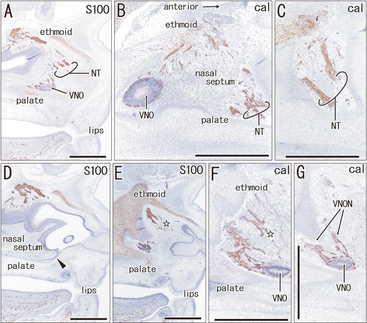
Fig. 1 The terminal nerve and nerves to the vomeronasal organ (VNON) are closely located in a specimen of crown rump length 45 mm (9 weeks). Sagittal sections. Immunohistochemistry of S100 protein (S100) (A, D, E) or calretinin (cal) (B, C, F, G). Panels A and D display the lateral planes in the figure, while panels B, C, F, and G display the medial planes. Thus, the upper or lower raw of panels exhibits a different side from the lateral to the medial, respectively. Intervals between panels are 0.2 mm (A–B), 0.1 mm (B–C), 0.2 mm (D–E), and 0.1 mm (E–F, F–G). On one side (panels A–C), the proximal course of the VNON was not cut, and we did not identify the distal course of the terminal nerve (NT) on the other side (panels D–G). Open stars in panels E and F indicate the most likely candidate of the terminal nerve. Arrowhead in panel D indicates an on-going fusion between the nasal septum and palate. In the specimen, the terminal nerve and VNON appeared to be closely located in the nasal septum. VNO, vomeronasal organ. Scale bars=1 mm (A–G).
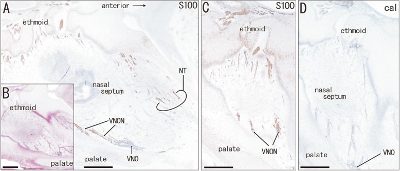
Fig. 2 The terminal nerve and nerves to the vomeronasal organ (VNON) are separated by a developing nasal septum in a specimen of crown rump length 110 mm (15 weeks). Sagittal sections. Immunohistochemistry of S100 protein (S100) (A, C) or calretinin (cal) (D). (B) Hematoxylin and eosin staining. Panels C and D display the vomeronasal organ (VNO) and VNON in the other side of panels A and B. Intervals between panels are 0.2 mm (A–B), 0.6 mm (B–C), and 0.1 mm (C–D). In panel A, the terminal nerve (NT) runs along the posterior aspect of the cartilaginous nasal bone, while the VNON run along the palate near the VNO. Thus, these nerves are separated by a developing nasal septum. Scale bars=1 mm (A–D).
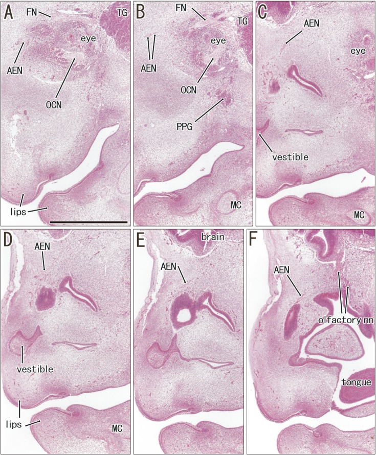
Fig. 3 Anterior ethmoidal nerve coming from the orbit in a specimen of crown rump length 27 mm (7 weeks). Sagittal sections. Hematoxylin and eosin staining. Panel A (or F) displays the most lateral (or medial) plane in the figure. Intervals between panels are 0.3 mm (A–B, B–C), 0.05 mm (C–D, D–E), and 0.1 mm (E–F). The anterior ethmoidal nerve (AEN) runs medially from the orbit (A–C), reaches the superolateral angle of the nasal cavity (D), and directs inferiorly along the mucosal membrane (E, F). Panels A–D were prepared at the same magnification (scale bar in panel A=1 mm). FN, frontal nerve; MC, Meckel's cartilage; OCN, oculomotor nerve; PPG, pterygopalatine ganglion; TG, trigeminal nerve ganglion.
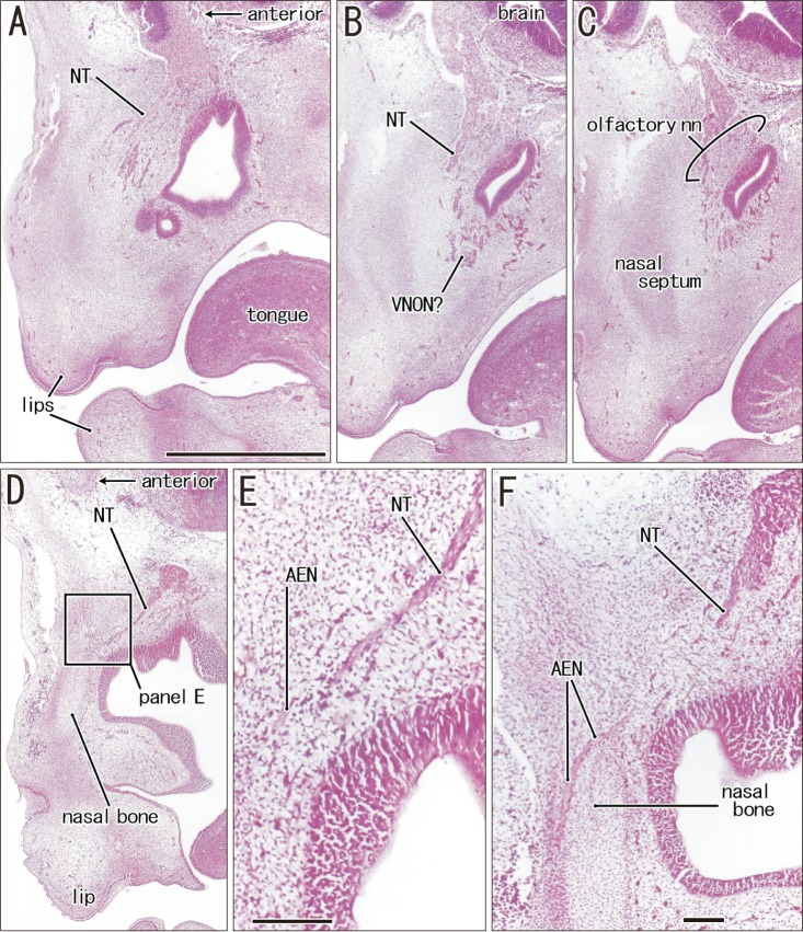
Fig. 4 Anterior ethmoidal nerve crossing superficially the terminal nerve in a specimen of crown rump length 27 mm (7 weeks). Sagittal sections. Hematoxylin and eosin staining. Panels A and F display the lateral sites, while panels B and C show planes near the midsagittal plane. Thus, the terminal nerve (NT) is seen bilaterally (A, D). Panel F is 0.2 mm medial to Fig. 3F. Panel E is the higher magnification view of a square in panel D. Intervals between panels are 0.2 mm (A–B), 0.1 mm (B–C, C–D, D–F). The terminal nerve appears to be still closely related to the nerves to the vomeronasal organ (VNON) candidate (VNON? in panel B). In the immediately superior side of the cartilaginous nasal bone, the anterior ethmoidal nerve (AEN) crosses superficially the terminal nerve without communication (E, F). Panels A–D were prepared at the same magnification. Scale bars=1 mm (A), 0.1 mm (E, F).
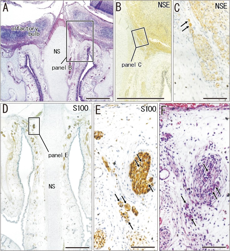
Fig. 5 Ganglion cells on the lateral surface of the crista galli of the ethmoid in a specimen of crown rump length 160 mm (18 weeks). Frontal sections. Panel A is 2.0 mm anterior to panel D. Panel B (or C) is a higher magnification view of a square in panel A (or B). Panel E is a higher magnification view of a square in panel D. Panel F is a section near panel E. Hematoxylin and eosin staining (A, F) or immunohistochemistry of neuron specific enolase (NSE) (B, C) and S100 protein (S100) (D, E). Ganglion cells on the crista galli are, 15–20 µm in diameter, positive for NSE (arrows in panels C), negative for S100 (arrows in panel E), and highly eosinophilic (arrows in panel F). Scale bars=1 mm (A, B, D), 0.1 mm (C, E, F). NS, nasal septum.
Reference
-
1. Johnston JB. The nervus terminalis in man and mammals. Anat Rec. 1914; 8:185–198.2. Larsell O. The nervus terminalis. Ann Otol Rhinol Laryngol. 1950; 59:414–438. PMID: 15426155.3. McCotter RE. A note on the course and distribution of the nervus terminalis in man. Anat Rec. 1915; 9:243–246.4. Pearson AA. The development of the nervus terminalis in man. J Comp Neurol. 1941; 75:39–66.5. Brookover C. The nervus terminalis in adult man. J Comp Neurol. 1914; 24:131–135.6. Fuller GN, Burger PC. Nervus terminalis (cranial nerve zero) in the adult human. Clin Neuropathol. 1990; 9:279–283. PMID: 2286018.7. Kim K, Patel L, Tobet S, King J, Rubin B, Stopa E. Gonadotropin-releasing hormone immunoreactivity in the adult and fetal human olfactory system. Brain Res. 1999; 826:220–229. PMID: 10224299.8. Müller F, O'rahilly R. Olfactory structures in staged human embryos. Cells Tissues Organs. 2004; 178:93–116. PMID: 15604533.9. Smith TD, Bhatnagar KP. The human vomeronasal organ. Part II: prenatal development. J Anat. 2000; 197:421–436. PMID: 11117628.10. Besli R, Saylam C, Veral A, Karl B, Ozek C. The existence of the vomeronasal organ in human beings. J Craniofac Surg. 2004; 15:730–735. PMID: 15346008.11. Demski LS. Terminal nerve complex. Acta Anat (Basel). 1993; 148:81–95. PMID: 8109200.12. Wirsig-Wiechmann CR, Wiechmann AF, Eisthen HL. What defines the nervus terminalis? Neurochemical, developmental, and anatomical criteria. Prog Brain Res. 2002; 141:45–58. PMID: 12508560.13. Bédard A, Parent A. Evidence of newly generated neurons in the human olfactory bulb. Brain Res Dev Brain Res. 2004; 151:159–168. PMID: 15246702.14. Bastianelli E, Polans AS, Hidaka H, Pochet R. Differential distribution of six calcium-binding proteins in the rat olfactory epithelium during postnatal development and adulthood. J Comp Neurol. 1995; 354:395–409. PMID: 7541806.15. Johnson EW. CaBPs and other immunohistochemical markers of the human vomeronasal system: a comparison with other mammals. Microsc Res Tech. 1998; 41:530–541. PMID: 9712200.16. Johnson EW. Immunocytochemical characteristics of cells and fibers in the nasal mucosa of young and adult macaques. Anat Rec. 2000; 259:215–228. PMID: 10820323.17. Malz CR, Knabe W, Kuhn HJ. Calretinin immunoreactivity in the prenatally developing olfactory systems of the tree shrew Tupaia belangeri. Anat Embryol (Berl). 2002; 205:83–97. PMID: 12021911.18. Katori Y, Jin ZW, Kawase T, Hong KH, Murakami G, Cho BH. Developmental changes in the distribution of calretinin-immunoreactive cells in human fetal nasal epithelium. Okajimas Folia Anat Jpn. 2010; 87:5–10. PMID: 20715566.19. Kim JH, Cho KH, Jin ZW, Murakami G, Abe H, Chai OH. Ganglion cardiacum or juxtaductal body of human fetuses. Anat Cell Biol. 2018; 51:266–273. PMID: 30637161.20. Cho KH, Kim JH, Murakami G, Abe H, Rodríguez-Vázquez JF, Chai OH. Nerve distribution in myocardium including the atrial ventricular septa in late stage human fetuses. Anat Cell Biol. 2019; 52:48–56. PMID: 30984452.21. Patron V, Berkaoui J, Jankowski R, Lechapt-Zalcman E, Moreau S, Hitier M. The forgotten foramina: a study of the anterior cribriform plate. Surg Radiol Anat. 2015; 37:835–840. PMID: 25823692.22. Bhatnagar KP, Kallen FC. Morphology of the nasal cavities and associated structures in Artibeus jamaicensis and Myotis lucifugus. Am J Anat. 1974; 139:167–189. PMID: 4812217.23. Huber GC, Guild SR. Observations on the peripheral distribution of the nervus terminalis in mammalia. Anat Rec. 1913; 7:253–272.24. Peña-Melián Á, Cabello-de la Rosa JP, Gallardo-Alcañiz MJ, Vaamonde-Gamo J, Relea-Calatayud F, González-López L, Villanueva-Anguita P, Flores-Cuadrado A, Saiz-Sánchez D, Martínez-Marcos A. Cranial pair 0: the nervus terminalis. Anat Rec (Hoboken). 2019; 302:394–404. PMID: 29663690.25. Dare AO, Balos LL, Grand W. Neural-dural transition at the medial anterior cranial base: an anatomical and histological study with clinical applications. J Neurosurg. 2003; 99:362–365. PMID: 12924711.
- Full Text Links
-
- Actions
-
Cited
- CITED
-
- Close
- Share
-
- Similar articles
-
- Histochemical Localization of NADPH-Diaphorase in the Rat Vomeronasal Organ
- A Case of Combined Nervus Showing Features of Common Blue Nervus and Dysplastic Nervus
- Histochemical Characterization of the Lectin-binding Sites in the Equine Vomeronasal Organ
- Morphological Study of the Ventriculus Terminalis in the Human Fetal Spinal Cord
- Developmental Changes of Glial Fibrillary Acidic Protein (GFAP) and Proliferating Cell Nuclear Antigen (PCNA) Immunoreactivity of the Ependyma lining the Central Canal and Ventriculus Terminalis in the Human Fetus
