Disruption of the Tff1 gene in mice using CRISPR/Cas9 promotes body weight reduction and gastric tumorigenesis
- Affiliations
-
- 1Department of Biochemistry, College of Life Science and Biotechnology and Yonsei Laboratory Animal Research Center, Yonsei University, Seoul, Korea. hwl@yonsei.ac.kr, jhlee13@gmail.com
- 2Severance Biomedical Science Institute, Brain Korea 21 PLUS Project for Medical Science, Yonsei University College of Medicine, Seoul, Korea. KITAEK@yuhs.ac
- KMID: 2459303
- DOI: http://doi.org/10.5625/lar.2018.34.4.257
Abstract
- Trefoil factor 1 (TFF1, also known as pS2) is strongly expressed in the gastrointestinal mucosa and plays a critical role in the differentiation of gastric glands. Since approximately 50% of all human gastric cancers are associated with decreased TFF1 expression, it is considered a tumor suppressor gene. TFF1 deficiency in mice results in histological changes in the antral and pyloric gastric mucosa, with severe hyperplasia and dysplasia of epithelial cells, resulting in the development of antropyloric adenoma. Here, we generated TFF1-knockout (KO) mice, without a neomycin resistant (NeoR) cassette, using the clustered regularly interspaced short palindromic repeats/CRISPR-associated nuclease 9 (CRSIPR/Cas9) system. Though our TFF1-KO mice showed phenotypes very similar to the previous embryonic stem (ES)-cell-based KO mice, they differed from the previous reports in that a reduction in body weight was observed in males. These results demonstrate that these newly established TFF1-KO mice are useful tools for investigating genetic and environmental factors influencing gastric cancer, without the effects of artificial gene insertion. Furthermore, these findings suggest a novel hypothesis that TFF1 expression influences gender differences.
Keyword
MeSH Terms
Figure
-
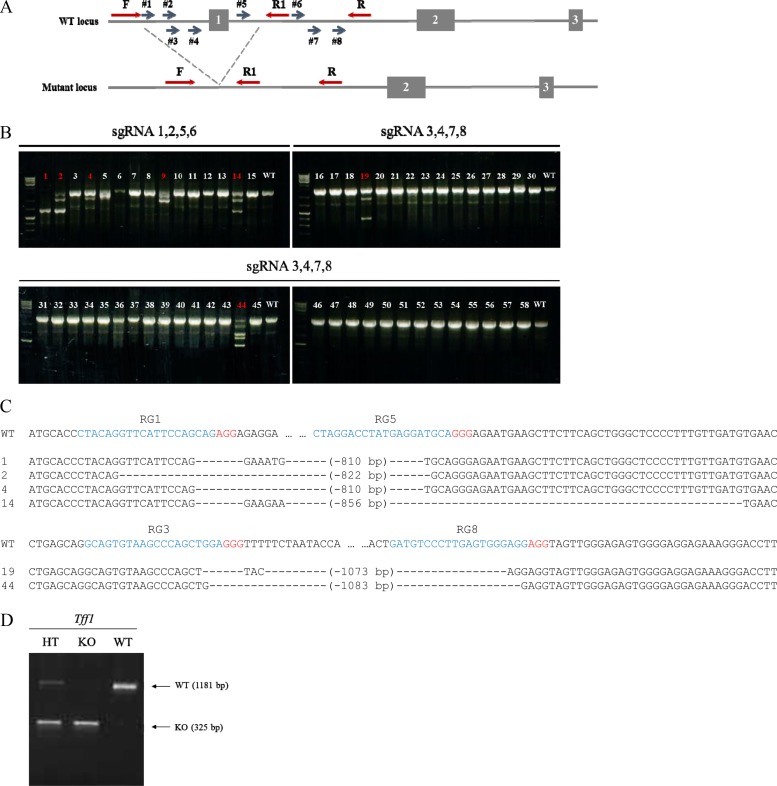
Figure 1 (A) CRISPR/Cas9 targeting strategy to generate Tff1-KO mice. Blue arrows #1–8 indicate the location and orientation of sgRNAs designed to target specific sites. Exons and introns are indicated with boxes and lines, respectively. PCR primers (F, forward; R, reverse) for genotyping are denoted by red arrows. (B) PCR screening results for newborns edited by Cas9 with sgRNAs 1, 2, 5, and 6 (combination 1) or 3, 4, 7, and 8 (combination 2). Mice carrying deletion mutations are highlighted in red. (C) Sequencing results for candidate newborns from combination 1 (upper panel) and 2 (bottom panel). The protospacer adjacent motif (PAM) and target sequences of sgRNAs are shown in red and blue, respectively. The symbol, “-”, indicates a single-nucleotide deletion. (D) PCR genotyping results for heterozygous (HT), homozygous (KO), and wild-type (WT) mice. Each arrow indicates WT-(1181 bp) and KO-specific (325 bp) bands.
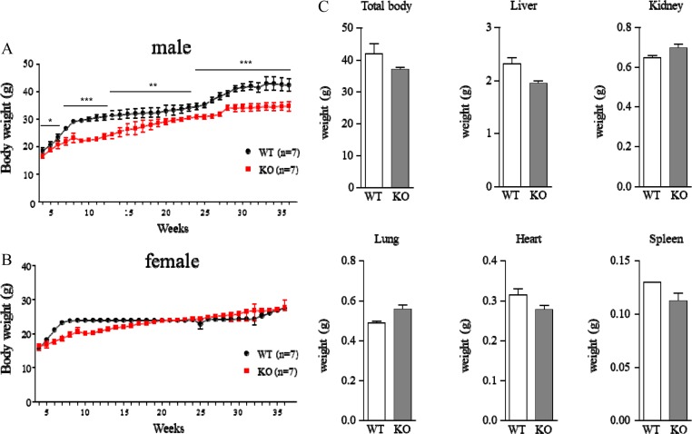
Figure 2 (A–B) Body weight changes in WT (black, n=7) and Tff1-KO (red, n=7) mice. Total body weights of male (A) and female (B) mice were measured once a week from 4 weeks to 36 weeks. Data are presented as mean±SEM (Student's t-test, *P<0.05, **P<0.01, ***P<0.001). (C) Mean weights (g) of total body, liver, kidney, lung, heart, and spleen in male WT and Tff1-KO mice after sacrifice. Data are presented as mean±SEM (WT, n=2; KO, n=7).
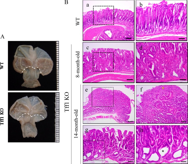
Figure 3 (A) Representative macroscopic images of WT (upper panel) and Tff1-KO mice (lower panel) at 8 months. The dotted line indicates the boundaries between the fundus and the antrum. (B) Representative H&E images of WT (a, b), 8-month-old (c, d) and 14-month-old Tff1-KO mice (e-h). Higher magnification views of images in the dotted boxes in the left panels are shown on the right. Hyperproliferation of the gastric epithelium and infiltration of immune cells are observed in the antrum of Tff1-KO mice (c, d). Adenoma is observed in the antrum region of Tff1-KO mice (e, f). Yellow asterisk indicates the tumor region (f). Mild lesions are observed in 14-month-old Tff1-KO mice (g, h). Black asterisk indicates cystic dilatation (h). Bars: 200 µm (e); 100 µm (a, c, f, g); 40 µm (b, d, h).
Reference
-
1. Roder DM. The epidemiology of gastric cancer. Gastric Cancer. 2002; 5(Suppl 1):5–11. PMID: 12772880.
Article2. Kamangar F, Dores GM, Anderson WF. Patterns of cancer incidence, mortality, and prevalence across five continents: defining priorities to reduce cancer disparities in different geographic regions of the world. J Clin Oncol. 2006; 24(14):2137–2150. PMID: 16682732.
Article3. Muoz N, Franceschi S. Epidemiology of gastric cancer and perspectives for prevention. Salud Publica Mex. 1997; 39(4):318–330. PMID: 9337564.
Article4. Hohenberger P, Gretschel S. Gastric cancer. Lancet. 2003; 362(9380):305–315. PMID: 12892963.5. Shi SQ, Cai JT, Yang JM. Expression of trefoil factors 1 and 2 in precancerous condition and gastric cancer. World J Gastroenterol. 2006; 12(19):3119–3122. PMID: 16718800.
Article6. Jakowlew SB, Breathnach R, Jeltsch JM, Masiakowski P, Chambon P. Sequence of the pS2 mRNA induced by estrogen in the human breast cancer cell line MCF-7. Nucleic Acids Res. 1984; 12(6):2861–2878. PMID: 6324130.
Article7. Xiao P, Ling H, Lan G, Liu J, Hu H, Yang R. Trefoil factors: Gastrointestinal-specific proteins associated with gastric cancer. Clin Chim Acta. 2015; 450:127–134. PMID: 26265233.
Article8. Lefebvre O, Chenard MP, Masson R, Linares J, Dierich A, LeMeur M, Wendling C, Tomasetto C, Chambon P, Rio MC. Gastric mucosa abnormalities and tumorigenesis in mice lacking the pS2 trefoil protein. Science. 1996; 274(5285):259–262. PMID: 8824193.
Article9. Ribieras S, Tomasetto C, Rio MC. The pS2/TFF1 trefoil factor, from basic research to clinical applications. Biochim Biophys Acta. 1998; 1378(1):F61–F77. PMID: 9739760.
Article10. Rio MC, Bellocq JP, Daniel JY, Tomasetto C, Lathe R, Chenard MP, Batzenschlager A, Chambon P. Breast cancer-associated pS2 protein: synthesis and secretion by normal stomach mucosa. Science. 1988; 241(4866):705–708. PMID: 3041593.
Article11. Wright AV, Nuez JK, Doudna JA. Biology and Applications of CRISPR Systems: Harnessing Nature's Toolbox for Genome Engineering. Cell. 2016; 164(1-2):29–44. PMID: 26771484.
Article12. Lee JH, Park JH, Nam TW, Seo SM, Kim JY, Lee HK, Han JH, Park SY, Choi YK, Lee HW. Differences between immunodeficient mice generated by classical gene targeting and CRISPR/Cas9-mediated gene knockout. Transgenic Res. 2018; 27(3):241–251. PMID: 29594927.
Article13. Roh JI, Lee J, Park SU, Kang YS, Lee J, Oh AR, Choi DJ, Cha JY, Lee HW. CRISPR-Cas9-mediated generation of obese and diabetic mouse models. Exp Anim. 2018; 67(2):229–237. PMID: 29343656.
Article14. Sung YH, Kim JM, Kim HT, Lee J, Jeon J, Jin Y, Choi JH, Ban YH, Ha SJ, Kim CH, Lee HW, Kim JS. Highly efficient gene knockout in mice and zebrafish with RNA-guided endonucleases. Genome Res. 2014; 24(1):125–131. PMID: 24253447.
Article15. Henry JA, Bennett MK, Piggott NH, Levett DL, May FE, Westley BR. Expression of the pNR-2/pS2 protein in diverse human epithelial tumours. Br J Cancer. 1991; 64(4):677–682. PMID: 1911216.
Article16. Luqmani Y, Bennett C, Paterson I, Corbishley CM, Rio MC, Chambon P, Ryall G. Expression of the pS2 gene in normal, benign and neoplastic human stomach. Int J Cancer. 1989; 44(5):806–812. PMID: 2583860.17. Machado JC, Carneiro F, Ribeiro P, Blin N, Sobrinho-Simes M. pS2 protein expression in gastric carcinoma. An immunohistochemical and immunoradiometric study. Eur J Cancer. 1996; 32A(9):1585–1590. PMID: 8911122.
Article18. Theisinger B, Welter C, Seitz G, Rio MC, Lathe R, Chambon P, Blin N. Expression of the breast cancer associated gene pS2 and the pancreatic spasmolytic polypeptide gene (hSP) in diffuse type of stomach carcinoma. Eur J Cancer. 1991; 27(6):770–773. PMID: 1829922.
Article19. Wanebo HJ, Kennedy BJ, Chmiel J, Steele G Jr, Winchester D, Osteen R. Cancer of the stomach. A patient care study by the American College of Surgeons. Ann Surg. 1993; 218(5):583–592. PMID: 8239772.20. Everett SM, Axon AT. Early gastric cancer in Europe. Gut. 1997; 41(2):142–150. PMID: 9301490.
Article21. Saukkonen K, Tomasetto C, Narko K, Rio MC, Ristimki A. Cyclooxygenase-2 expression and effect of celecoxib in gastric adenomas of trefoil factor 1-deficient mice. Cancer Res. 2003; 63(12):3032–3036. PMID: 12810622.22. Blum D, Stene GB, Solheim TS, Fayers P, Hjermstad MJ, Baracos VE, Fearon K, Strasser F, Kaasa S. Euro-Impact. Validation of the Consensus-Definition for Cancer Cachexia and evaluation of a classification model--a study based on data from an international multicentre project (EPCRC-CSA). Ann Oncol. 2014; 25(8):1635–1642. PMID: 24562443.
Article23. Artelt P, Grannemann R, Stocking C, Friel J, Bartsch J, Hauser H. The prokaryotic neomycin-resistance-encoding gene acts as a transcriptional silencer in eukaryotic cells. Gene. 1991; 99(2):249–254. PMID: 1850711.
Article
- Full Text Links
-
- Actions
-
Cited
- CITED
-
- Close
- Share
-
- Similar articles
-
- CRISPR-Cas9 system in autosomal dominant polycystic kidney disease: a comprehensive review
- Construction of a CRISPR/Cas9-Mediated Genome Editing System in Lentinula edodes
- Two base pair deletion in IL2 receptor γ gene in NOD/SCID mice induces a highly severe immunodeficiency
- Genome editing: the road of CRISPR/Cas9 from bench to clinic
- Applications of CRISPR/Cas9 for Gene Editing in Hereditary Movement Disorders
