Tuberc Respir Dis.
2019 Oct;82(4):269-276. 10.4046/trd.2018.0090.
Korean Guidelines for Diagnosis and Management of Interstitial Lung Diseases: Part 1. Introduction
- Park SW

- Baek AR
- Lee HL
- Jeong SW
- Yang SH
- Kim YH
- Chung MP
- on behalf of the Korean Interstitial Lung Diseases Study Group
- Affiliations
-
- 1Division of Respiratory and Allergy, Department of Internal Medicine, Soonchunhyang University Bucheon Hospital, Soonchunhyang University College of Medicine, Bucheon, Korea.
- 2Division of Pulmonary and Critical Care Medicine, Department of Internal Medicine, Inha University College of Medicine, Incheon, Korea.
- 3Department of Internal Medicine, Gachon University Gil Medical Center, Incheon, Korea.
- 4Division of Pulmonary and Critical Care Medicine, Department of Internal Medicine, Wonkwang University School of Medicine, Iksan, Korea.
- 5Division of Allergy and Pulmonology, Department of Internal Medicine, Bucheon St. Mary's Hospital, College of Medicine, The Catholic University of Korea, Bucheon, Korea.
- 6Division of Pulmonary and Critical Care Medicine, Department of Medicine, Samsung Medical Center, Sungkyunkwan University School of Medicine, Seoul, Korea. mpchung@skku.edu
- KMID: 2459050
- DOI: http://doi.org/10.4046/trd.2018.0090
Abstract
- Idiopathic interstitial pneumonia (IIP) is a histologically identifiable pulmonary disease without a known cause that usually infiltrates the lung interstitium. IIP is largely classified into idiopathic pulmonary fibrosis, idiopathic non-specific interstitial pneumonia, respiratory bronchiolitis-interstitial lung disease (ILD), cryptogenic organizing pneumonia, desquamative interstitial pneumonia, and acute interstitial pneumonia. Each of these diseases has a different prognosis and requires specific treatment, and a multidisciplinary approach that combines chest high-resolution computed tomography (HRCT), histological findings, and clinical findings is necessary for their diagnosis. Diagnosis of IIP is made based on clinical presentation, chest HRCT findings, results of pulmonary function tests, and histological findings. For histological diagnosis, video-assisted thoracoscopic biopsy and transbronchial lung biopsy are used. In order to identify ILD associated with connective tissue disease, autoimmune antibody tests may also be necessary. Many biomarkers associated with disease prognosis have been recently discovered, and future research on their clinical significance is necessary. The diagnosis of ILD is difficult because patterns of ILD are both complicated and variable. Therefore, as with other diseases, accurate history taking and meticulous physical examination are crucial.
MeSH Terms
Figure
-
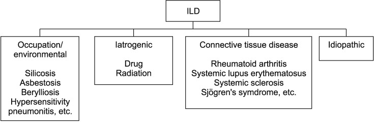
Figure 1 Classification of interstitial lung disease (ILD). ILD can be primarily classified with and without known causes. Known etiologies of ILD were occupational or environmental exposures, drugs, radiation, connective tissue diseases and so on.
Reference
-
1. Travis WD, Costabel U, Hansell DM, King TE Jr, Lynch DA, Nicholson AG, et al. An official American Thoracic Society/European Respiratory Society statement: Update of the international multidisciplinary classification of the idiopathic interstitial pneumonias. Am J Respir Crit Care Med. 2013; 188:733–748. PMID: 24032382.
Article2. American Thoracic Society. European Respiratory Society. American Thoracic Society/European Respiratory Society International Multidisciplinary Consensus Classification of the Idiopathic Interstitial Pneumonias. This joint statement of the American Thoracic Society (ATS), and the European Respiratory Society (ERS) was adopted by the ATS board of directors, June 2001 and by the ERS Executive Committee, June 2001. Am J Respir Crit Care Med. 2002; 165:277–304. PMID: 11790668.3. Corte TJ, Copley SJ, Desai SR, Zappala CJ, Hansell DM, Nicholson AG, et al. Significance of connective tissue disease features in idiopathic interstitial pneumonia. Eur Respir J. 2012; 39:661–668. PMID: 21920896.
Article4. Nogee LM, Dunbar AE 3rd, Wert SE, Askin F, Hamvas A, Whitsett JA. A mutation in the surfactant protein C gene associated with familial interstitial lung disease. N Engl J Med. 2001; 344:573–579. PMID: 11207353.
Article5. Hodgson U, Laitinen T, Tukiainen P. Nationwide prevalence of sporadic and familial idiopathic pulmonary fibrosis: evidence of founder effect among multiplex families in Finland. Thorax. 2002; 57:338–342. PMID: 11923553.
Article6. Seibold MA, Wise AL, Speer MC, Steele MP, Brown KK, Loyd JE, et al. A common MUC5B promoter polymorphism and pulmonary fibrosis. N Engl J Med. 2011; 364:1503–1512. PMID: 21506741.7. Reddy TL, Tominaga M, Hansell DM, von der Thusen J, Rassl D, Parfrey H, et al. Pleuroparenchymal fibroelastosis: a spectrum of histopathological and imaging phenotypes. Eur Respir J. 2012; 40:377–385. PMID: 22441748.
Article8. Raghu G, Collard HR, Egan JJ, Martinez FJ, Behr J, Brown KK, et al. An official ATS/ERS/JRS/ALAT statement: idiopathic pulmonary fibrosis: evidence-based guidelines for diagnosis and management. Am J Respir Crit Care Med. 2011; 183:788–824. PMID: 21471066.9. Tomassetti S, Wells AU, Costabel U, Cavazza A, Colby TV, Rossi G, et al. Bronchoscopic lung cryobiopsy increases diagnostic confidence in the multidisciplinary diagnosis of idiopathic pulmonary fibrosis. Am J Respir Crit Care Med. 2016; 193:745–752. PMID: 26562389.
Article
- Full Text Links
-
- Actions
-
Cited
- CITED
-
- Close
- Share
-
- Similar articles
-
- Update in Diagnosis of Idiopathic Pulmonary Fibrosis and Interstitial Lung Abnormality
- Korean Guidelines for Diagnosis and Management of Interstitial Lung Diseases: Part 5. Connective Tissue Disease Associated Interstitial Lung Disease
- New Era of Management Concept on Pulmonary Fibrosis with Revisiting Framework of Interstitial Lung Diseases
- Korean Guidelines for Diagnosis and Management of Interstitial Lung Diseases: Connective Tissue Disease Associated Interstitial Lung Disease
- Idiopathic Interstitial Pneumonias: Radiologic Findings
