J Korean Soc Radiol.
2019 Sep;80(5):930-941. 10.3348/jksr.2019.80.5.930.
Relationship between Epicardial Fat Accumulation and Left Atrial Reverse Remodeling after Catheter Ablation of Atrial Fibrillation
- Affiliations
-
- 1Department of Radiology, Korea University Anam Hospital, Seoul, Korea. sungho.hwng@gmail.com
- 2Division of Cardiology, Department of Internal Medicine, Korea University Anam Hospital, Seoul, Korea.
- KMID: 2459018
- DOI: http://doi.org/10.3348/jksr.2019.80.5.930
Abstract
- PURPOSE
To demonstrate the relationship between epicardial fat accumulation and left atrial reverse remodeling by cardiac multi-detector CT (MDCT) after catheter ablation of atrial fibrillation (AF).
MATERIALS AND METHODS
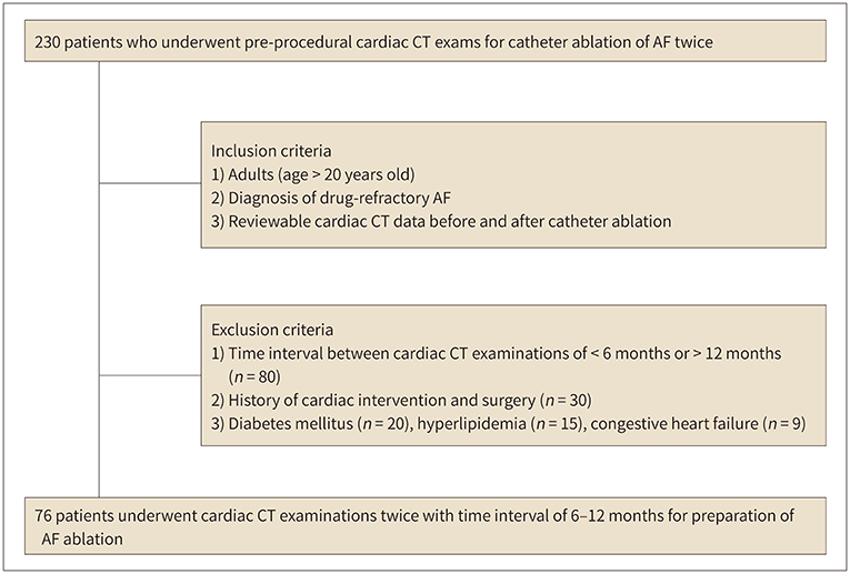
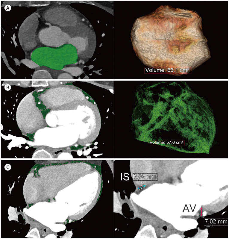
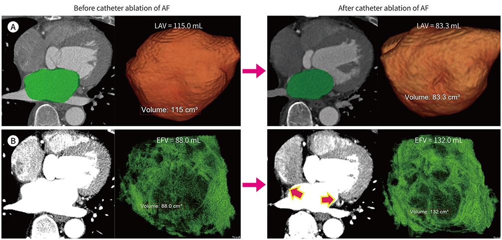
Seventy-six patients underwent cardiac MDCT before and after catheter ablation of AF. Left atrial volume (LAV) and epicardial fat volume (EFV) were measured. LAV and EFV before and after catheter ablation of AF were respectively compared and the change percentages (CPs) were evaluated.
RESULTS
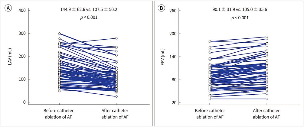
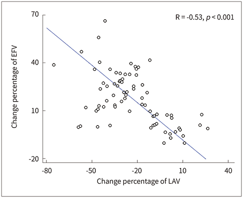
The LAV after catheter ablation of AF was significantly less than the baseline LAV (107.5 ± 50.2 mL vs. 144.9 ± 62.6 mL, p < 0.001). The EFV after catheter ablation of AF was significantly greater than the baseline EFV (105.0 ± 35.6 mL vs. 90.1 ± 31.9 mL, p < 0.001). Mean CPs of LAV and EFV were −23.3% ± 20.8% and 15.9% ± 20.9%, respectively. There was a significantly negative relationship between the CPs of LAV and EFV (R = −0.53, p < 0.001).
CONCLUSION
Catheter ablation of AF may result in a reduction in LAV and an increase in EFV. Left atrial reverse remodeling with a reduction in LAV may be associated with epicardial fat accumulation in patients who undergo catheter ablation of AF.
MeSH Terms
Figure
Reference
-
1. Fuster V, Rydén LE, Cannom DS, Crijns HJ, Curtis AB, Ellenbogen KA, et al. ACC/AHA/ESC 2006 guidelines for the management of patients with atrial fibrillation: a report of the American College of Cardiology/American Heart Association Task Force on Practice Guidelines and the European Society of Cardiology Committee for Practice Guidelines (Writing Committee to Revise the 2001 Guidelines for the Management of Patients With Atrial Fibrillation): developed in collaboration with the European Heart Rhythm Association and the Heart Rhythm Society. Circulation. 2006; 114:e257–e354.2. Goette A, Honeycutt C, Langberg JJ. Electrical remodeling in atrial fibrillation. Time course and mechanisms. Circulation. 1996; 94:2968–2974.3. Nattel S, Burstein B, Dobrev D. Atrial remodeling and atrial fibrillation: mechanisms and implications. Circ Arrhythm Electrophysiol. 2008; 1:62–73.4. Camm AJ, Lip GY, De Caterina R, Savelieva I, Atar D, Hohnloser SH, et al. 2012 focused update of the ESC guidelines for the management of atrial fibrillation: an update of the 2010 ESC guidelines for the management of atrial fibrillation. Developed with the special contribution of the European Heart Rhythm Association. Eur Heart J. 2012; 33:2719–2747.5. Arbelo E, Brugada J, Blomström-Lundqvist C, Laroche C, Kautzner J, Pokushalov E, et al. Contemporary management of patients undergoing atrial fibrillation ablation: in-hospital and 1-year follow-up findings from the ESC-EHRA atrial fibrillation ablation long-term registry. Eur Heart J. 2017; 38:1303–1316.
Article6. Jayam VK, Dong J, Vasamreddy CR, Lickfett L, Kato R, Dickfeld T, et al. Atrial volume reduction following catheter ablation of atrial fibrillation and relation to reduction in pulmonary vein size: an evaluation using magnetic resonance angiography. J Interv Card Electrophysiol. 2005; 13:107–114.
Article7. Jeevanantham V, Ntim W, Navaneethan SD, Shah S, Johnson AC, Hall B, et al. Meta-analysis of the effect of radiofrequency catheter ablation on left atrial size, volumes and function in patients with atrial fibrillation. Am J Cardiol. 2010; 105:1317–1326.
Article8. Reant P, Lafitte S, Jaïs P, Serri K, Weerasooriya R, Hocini M, et al. Reverse remodeling of the left cardiac chambers after catheter ablation after 1 year in a series of patients with isolated atrial fibrillation. Circulation. 2005; 112:2896–2903.
Article9. Iacobellis G, Camarena V, Sant DW, Wang G. Human epicardial fat expresses glucagon-like peptide 1 and 2 receptors genes. Horm Metab Res. 2017; 49:625–630.
Article10. Chang TY, Hsu CY, Chiu CC, Chou RH, Huang HL, Huang CC, et al. Association between echocardiographic epicardial fat thickness and circulating endothelial progenitor cell level in patients with stable angina pectoris. Clin Cardiol. 2017; 40:697–703.
Article11. Nishio S, Kusunose K, Yamada H, Hirata Y, Ise T, Yamaguchi K, et al. Echocardiographic epicardial adipose tissue thickness is associated with symptomatic coronary vasospasm during provocative testing. J Am Soc Echocardiogr. 2017; 30:1021–1027.e1.
Article12. Wong CX, Ganesan AN, Selvanayagam JB. Epicardial fat and atrial fibrillation: current evidence, potential mechanisms, clinical implications, and future directions. Eur Heart J. 2017; 38:1294–1302.
Article13. Batal O, Schoenhagen P, Shao M, Ayyad AE, Van Wagoner DR, Halliburton SS, et al. Left atrial epicardial adiposity and atrial fibrillation. Circ Arrhythm Electrophysiol. 2010; 3:230–236.
Article14. Nakanishi R, Rajani R, Cheng VY, Gransar H, Nakazato R, Shmilovich H, et al. Increase in epicardial fat volume is associated with greater coronary artery calcification progression in subjects at intermediate risk by coronary calcium score: a serial study using non-contrast cardiac CT. Atherosclerosis. 2011; 218:363–368.
Article15. Iacobellis G, Singh N, Wharton S, Sharma AM. Substantial changes in epicardial fat thickness after weight loss in severely obese subjects. Obesity (Silver Spring). 2008; 16:1693–1697.
Article16. Nagashima K, Okumura Y, Watanabe I, Nakai T, Ohkubo K, Kofune T, et al. Association between epicardial adipose tissue volumes on 3-dimensional reconstructed CT images and recurrence of atrial fibrillation after catheter ablation. Circ J. 2011; 75:2559–2565.
Article17. Stojanovska J, Cronin P, Patel S, Gross BH, Oral H, Chughtai K, et al. Reference normal absolute and indexed values from ECG-gated MDCT: left atrial volume, function, and diameter. AJR Am J Roentgenol. 2011; 197:631–637.
Article18. Vorre MM, Abdulla J. Diagnostic accuracy and radiation dose of CT coronary angiography in atrial fibrillation: systematic review and meta-analysis. Radiology. 2013; 267:376–386.
Article19. Gweon HM, Kim SJ, Kim TH, Lee SM, Hong YJ, Rim SJ. Evaluation of left atrial volumes using multidetector computed tomography: comparison with echocardiography. Korean J Radiol. 2010; 11:286–294.
Article20. Park HE, Choi SY, Kim HS, Kim MK, Cho SH, Oh BH. Epicardial fat reflects arterial stiffness: assessment using 256-slice multidetector coronary computed tomography and cardio-ankle vascular index. J Atheroscler Thromb. 2012; 19:570–576.
Article21. Shrout PE, Fleiss JL. Intraclass correlations: uses in assessing rater reliability. Psychol Bull. 1979; 86:420–428.
Article22. Beukema WP, Elvan A, Sie HT, Misier AR, Wellens HJ. Successful radiofrequency ablation in patients with previous atrial fibrillation results in a significant decrease in left atrial size. Circulation. 2005; 112:2089–2095.
Article23. Stojanovska J, Kazerooni EA, Sinno M, Gross BH, Watcharotone K, Patel S, et al. Increased epicardial fat is independently associated with the presence and chronicity of atrial fibrillation and radiofrequency ablation outcome. Eur Radiol. 2015; 25:2298–2309.
Article24. Nakazato R, Rajani R, Cheng VY, Shmilovich H, Nakanishi R, Otaki Y, et al. Weight change modulates epicardial fat burden: a 4-year serial study with non-contrast computed tomography. Atherosclerosis. 2012; 220:139–144.
Article25. Wong CX, Abed HS, Molaee P, Nelson AJ, Brooks AG, Sharma G, et al. Pericardial fat is associated with atrial fibrillation severity and ablation outcome. J Am Coll Cardiol. 2011; 57:1745–1751.
Article26. Fox CS, Gona P, Hoffmann U, Porter SA, Salton CJ, Massaro JM, et al. Pericardial fat, intrathoracic fat, and measures of left ventricular structure and function: the Framingham Heart Study. Circulation. 2009; 119:1586–1591.
- Full Text Links
- Actions
-
Cited
- CITED
-
- Close
- Share
- Similar articles
-
- Controlled Atrial Fibrillation after Pulmonary Vein Stenting
- A Case of Successful Ablation of Right-Sided Accessory Pathway during Atrial Fibrillation
- Underdevelopment of Left Atrial Appendage
- The Mechanism of and Preventive Therapy for Stroke in Patients with Atrial Fibrillation
- The Impact of the CHAâ‚‚DSâ‚‚-VASc Score on Recurrence of Atrial Fibrillation after a Single Catheter Ablation and Atrial Remodeling in Patients with Non-Valvular Atrial Fibrillation

