Dental students' ability to detect maxillary sinus abnormalities: A comparison between panoramic radiography and cone-beam computed tomography
- Affiliations
-
- 1Division of Oral Radiology, Department of Oral Diagnosis, Piracicaba Dental School, State University of Campinas, Piracicaba, São Paulo, Brazil. lucaslopesrosado@gmail.com
- 2Department of Dentistry, Federal University of Juiz de Fora Governador Valadares Campus, Governador Valadares, Minas Gerais, Brazil.
- 3Division of Oral Diagnosis, Department of Dentistry, Federal University of Juiz de Fora Governador Valadares Campus, Governador Valadares, Minas Gerais, Brazil.
- 4Division of Endodontics, Department of Dentistry, Federal University of Juiz de Fora Governador Valadares Advanced Campus, Governador Valadares, Minas Gerais, Brazil.
- KMID: 2458367
- DOI: http://doi.org/10.5624/isd.2019.49.3.191
Abstract
- PURPOSE
To compare the diagnostic ability of undergraduate dental students to detect maxillary sinus abnormalities in panoramic radiographs (PR) and cone-beam computed tomography (CBCT).
MATERIALS AND METHODS
This was a retrospective study based on the evaluation of PR and CBCT images. A pilot study was conducted to determine the number of students eligible to participate in the study. The images were evaluated by 2 students, and 280 maxillary sinuses were assessed using the following categories: normal, mucosal thickening, sinus polyp, antral pseudocyst, nonspecific opacification, periostitis, antrolith, and antrolith associated with mucosal thickening. The reference standard was established by the consensus of 2 oral radiologists based on the CBCT images. The kappa test, receiver operating characteristic curves, and 1-way analysis of variance with the Tukey-Kramer post-hoc test were employed.
RESULTS
Intraobserver and interobserver reliability showed agreement ranging from substantial (0.809) to almost perfect (0.922). The agreement between the students' evaluations and the reference standard was reasonable (0.258) for PR and substantial (0.692) for CBCT. Comparisons of values of sensitivity, specificity, and accuracy showed that CBCT was significantly better (P<0.05).
CONCLUSION
CBCT was better than PR for the detection of maxillary sinus abnormalities by dental students. However, CBCT should only be requested after a careful analysis of PR by students and more experienced professionals.
MeSH Terms
Figure
-
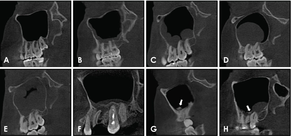
Fig. 1 Sagittal sections of cone-beam computed tomography show examples of maxillary sinus abnormalities. A. Normal. B. Mucosal thickening. C. Sinus polyp. D. Antral pseudocyst. E. Non-specific opacification. F. Periostitis. G. Antrolith. H. Antrolith associated with mucosal thickening. The arrows indicate an antrolith inside the maxillary sinus.
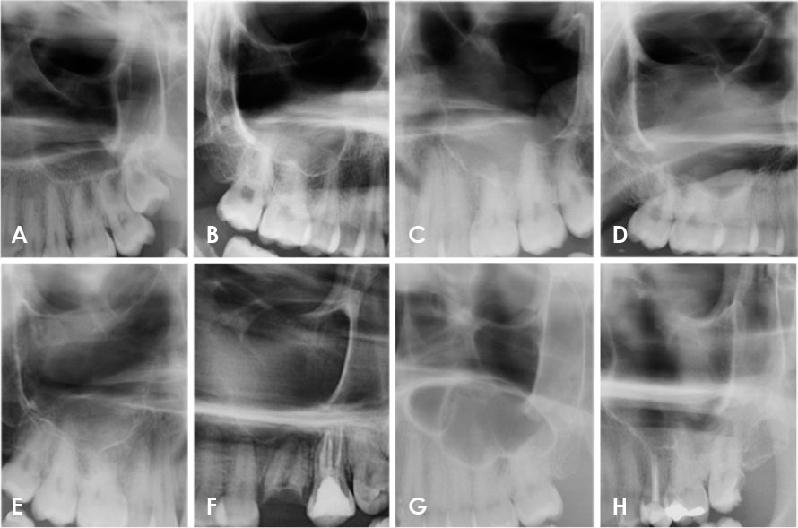
Fig. 2 Sections of cropped panoramic radiographs show examples of maxillary sinus abnormalities. A. Normal. B. Mucosal thickening. C. Sinus polyp. D. Antral pseudocyst. E. Non-specific opacification. F. Periostitis. G. Antrolith. H. Antrolith associated with mucosal thickening.

Fig. 3 Receiver operating characteristic curves. A. Panoramic radiographs. B. Cone-beam computed tomographic images.
Reference
-
1. Maillet M, Bowles WR, McClanahan SL, John MT, Ahmad M. Cone-beam computed tomography evaluation of maxillary sinusitis. J Endod. 2011; 37:753–757.
Article2. Maestre-Ferrín L, Galán-Gil S, Carrillo-Garcia C, Peñarrocha-Diago M. Radiographic findings in the maxillary sinus: comparison of panoramic radiography with computed tomography. Int J Oral Maxillofac Implants. 2011; 26:341–346.3. Nunes CA, Guedes OA, Alencar AH, Peters OA, Estrela CR, Estrela C. Evaluation of periapical lesions and their association with maxillary sinus abnormalities on cone-beam computed tomographic images. J Endod. 2016; 42:42–46.
Article4. Rege IC, Sousa TO, Leles CR, Mendonça EF. Occurrence of maxillary sinus abnormalities detected by cone beam CT in asymptomatic patients. BMC Oral Health. 2012; 12:30.
Article5. Tadinada A, Fung K, Thacker S, Mahdian M, Jadhav A, Schincaglia GP. Radiographic evaluation of the maxillary sinus prior to dental implant therapy: a comparison between two-dimensional and three-dimensional radiographic imaging. Imaging Sci Dent. 2015; 45:169–174.
Article6. Malina-Altzinger J, Damerau G, Grätz KW, Stadlinger PD. Evaluation of the maxillary sinus in panoramic radiography - a comparative study. Int J Implant Dent. 2015; 1:17.
Article7. Jaju PP, Jaju SP. Cone-beam computed tomography: time to move from ALARA to ALADA. Imaging Sci Dent. 2015; 45:263–265.
Article8. Dau M, Marciak P, Al-Nawas B, Staedt H, Alshiri A, Frerich B, et al. Evaluation of symptomatic maxillary sinus pathologies using panoramic radiography and cone beam computed tomography - influence of professional training. Int J Implant Dent. 2017; 3:13.
Article9. Landis JR, Koch GG. The measurement of observer agreement for categorical data. Biometrics. 1977; 33:159–174.
Article10. Jaju PP, Jaju SP. Clinical utility of dental cone-beam computed tomography: current perspectives. Clin Cosmet Investig Dent. 2014; 6:29–43.
Article11. Baciut M, Hedesiu M, Bran S, Jacobs R, Nackaerts O, Baciut G. Pre- and postoperative assessment of sinus grafting procedures using cone-beam computed tomography compared with panoramic radiographs. Clin Oral Implants Res. 2013; 24:512–516.
Article12. Shahbazian M, Jacobs R. Diagnostic value of 2D and 3D imaging in odontogenic maxillary sinusitis: a review of literature. J Oral Rehabil. 2012; 39:294–300.
Article13. Simuntis R, Kubilius R, Padervinskis E, Ryškienė S, Tušas P, Vaitkus S. Clinical efficacy of main radiological diagnostic methods for odontogenic maxillary sinusitis. Eur Arch Otorhinolaryngol. 2017; 274:3651–3658.
Article14. Shahbazian M, Vandewoude C, Wyatt J, Jacobs R. Comparative assessment of panoramic radiography and CBCT imaging for radiodiagnostics in the posterior maxilla. Clin Oral Investig. 2014; 18:293–300.
Article15. Phothikhun S, Suphanantachat S, Chuenchompoonut V, Nisapakultorn K. Cone-beam computed tomographic evidence of the association between periodontal bone loss and mucosal thickening of the maxillary sinus. J Periodontol. 2012; 83:557–564.
Article16. Venskutonis T, Plotino G, Tocci L, Gambarini G, Maminskas J, Juodzbalys G. Periapical and endodontic status scale based on periapical bone lesions and endodontic treatment quality evaluation using cone-beam computed tomography. J Endod. 2015; 41:190–196.
Article17. Nascimento EH, Pontual ML, Pontual AA, Freitas DQ, Perez DE, Ramos-Perez FM. Association between odontogenic conditions and maxillary sinus disease: a study using cone-beam computed tomography. J Endod. 2016; 42:1509–1515.
Article18. Gang TI, Huh KH, Yi WJ, Lee SS, Heo MS, Choi SC. The effect of radiographic imaging modalities and the observer's experience on postoperative maxillary cyst assessment. Imaging Sci Dent. 2014; 44:301–305.
Article19. Cho BH. Diagnostic performance of dental students in identifying mandibular condyle fractures by panoramic radiography and the usefulness of reference images. Imaging Sci Dent. 2011; 41:53–57.
Article20. Shintaku WH, Enciso R, Covington JS, Migliorati CA. Can dental students be taught to use dental radiographs for osteoporosis screening? J Dent Educ. 2013; 77:598–603.
Article21. Kratz RJ, Nguyen CT, Walton JN, MacDonald D. Dental students' interpretations of digital panoramic radiographs on completely edentate patients. J Dent Educ. 2018; 82:313–321.
Article22. Baghdady MT, Carnahan H, Lam EW, Woods NN. Integration of basic sciences and clinical sciences in oral radiology education for dental students. J Dent Educ. 2013; 77:757–763.
Article23. Baghdady MT, Carnahan H, Lam EW, Woods NN. Dental and dental hygiene students' diagnostic accuracy in oral radiology: effect of diagnostic strategy and instructional method. J Dent Educ. 2014; 78:1279–1285.
Article24. Turgeon DP, Lam EW. Influence of experience and training on dental students' examination performance regarding panoramic images. J Dent Educ. 2016; 80:156–164.
Article
- Full Text Links
-
- Actions
-
Cited
- CITED
-
- Close
- Share
-
- Similar articles
-
- Comparison of panoramic radiography and cone beam computed tomography for assessing the relationship between the maxillary sinus floor and maxillary molars
- Comparison of panoramic radiography and cone-beam computed tomography for assessing radiographic signs indicating root protrusion into the maxillary sinus
- The value of panoramic radiography in assessing maxillary sinus inflammation
- Assessment of maxillary third molars with panoramic radiography and cone-beam computed tomography
- Maxillary sinus septa: comparison between panoramic radiography and CBCT
