Ann Rehabil Med.
2019 Aug;43(4):524-529. 10.5535/arm.2019.43.4.524.
Ten-Year Follow-Up of Transcranial Magnetic Stimulation Study in a Patient With Congenital Mirror Movements: A Case Report
- Affiliations
-
- 1Department of Physical Medicine and Rehabilitation, Chonbuk National University Medical School, Jeonju, Korea. shpark0130@jbnu.ac.kr
- 2Research Institute of Clinical Medicine of Chonbuk National University–Biomedical Research Institute of Chonbuk National University Hospital, Jeonju, Korea.
- KMID: 2457761
- DOI: http://doi.org/10.5535/arm.2019.43.4.524
Abstract
- Most studies concerning congenital mirror movements (CMMs) have been focused on the motor organization in the distal hand muscles exclusively. To the best of our knowledge, there is no data on motor organization pattern of lower extremities, and a scarcity of data on the significance of forearm and arm muscles in CMMs. Here, we describe the case of a 19-year-old boy presenting mirror movements. In these terms, a 10-year transcranial magnetic stimulation study demonstrated that the motor organization pattern of the arm muscles was different from that of distal hand and forearm muscles even in the same upper extremity, and that the lower extremities showed the same pathways as healthy children. Moreover, in this case, an ipsilateral motor evoked potentials (MEPs) for distal hand muscles increased in amplitude with age, even though the intensity of mirror movements decreased. In the arm muscles, however, it was concluded that the contralateral MEPs increased in amplitude with age.
Keyword
MeSH Terms
Figure
-
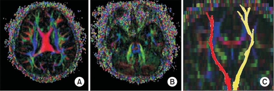
Fig. 1. Axial color-coded fractional anisotropy map demonstrating well-defined blue color corticospinal tract (CST) at the level of the cerebral cortex (A) and pons (B). The normal connectivity of whole CST was confirmed by diffusion tensor tractography (C).
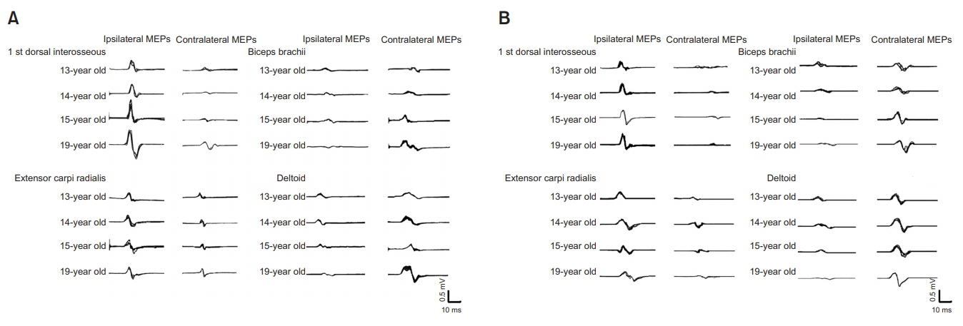
Fig. 2. Trend of motor evoked potentials (MEPs) with age, generated by transcranial magnetic stimulation at left motor cortex (A) and right motor cortex (B). For the 1st dorsal interosseous and extensor carpi radialis, ipsilateral MEPs increased in amplitude as the patient grew older. In contrast, the contralateral MEPs increased in amplitude for the biceps brachii and deltoid.
Reference
-
1. Maegaki Y, Seki A, Suzaki I, Sugihara S, Ogawa T, Amisaki T, et al. Congenital mirror movement: a study of functional MRI and transcranial magnetic stimulation. Dev Med Child Neurol. 2002; 44:838–43.
Article2. Park SH, Im KJ, Jo DS. Ipsilateral corticospinal projections in a patient with congenital mirror movements: a case report. J Korean Acad Rehabil Med. 2009; 33:502–5.3. Woods BT, Teuber HL. Mirror movements after childhood hemiparesis. Neurology. 1978; 28:1152–7.
Article4. Srour M, Riviere JB, Pham JM, Dube MP, Girard S, Morin S, et al. Mutations in DCC cause congenital mirror movements. Science. 2010; 328:592.5. Yook SW, Park SH, Ko MH, Seo JH. Motor evoked potentials of the upper extremities in healthy children. Ann Rehabil Med. 2011; 35:759–64.
Article6. Son SM, Park SH, Jo DS. Ipsilateral corticospinal projections in a patient with bilateral cortical malformation: a case report. J Korean Acad Rehabil Med. 2008; 32:582–5.7. Kim KW, Seo JH, Ko MH, Won YH, Park SH. A wide spectrum of axial mesodermal dysplasia complex with rhombencephalic anomaly: a case report. Ann Rehabil Med. 2016; 40:162–7.
Article8. Finger JH, Bronson RT, Harris B, Johnson K, Przyborski SA, Ackerman SL. The netrin 1 receptors Unc5h3 and Dcc are necessary at multiple choice points for the guidance of corticospinal tract axons. J Neurosci. 2002; 22:10346–56.9. Brandao P, Jovem C, Brasil-Neto JP, Tomaz C, Descoteaux M, Allam N. Congenital mirror movements: lack of decussation of pyramids. Brain. 2014; 137(Pt 8):e292.
- Full Text Links
-
- Actions
-
Cited
- CITED
-
- Close
- Share
-
- Similar articles
-
- Ipsilateral Corticospinal Projections in a Patient with Congenital Mirror Movements : A case report
- Motor Evoked Potential Study of Mirror Movements in a Patient with Klippel-Feil Syndrome
- Cortical Activation Related to Motor and Sensory Tasks in Congenital Mirror Movement using Functional MRI
- Neuromodulation for the Treatment of Tinnitus
- Motor Evoked Potentials in Masseter, and Anterior Belly of Digastric Induced by Transcranial Magnetic Stimulation
