Ann Dermatol.
2018 Aug;30(4):465-467. 10.5021/ad.2018.30.4.465.
Mucinous Nevus
- Affiliations
-
- 1Department of Dermatology, Ewha Womans University College of Medicine, Seoul, Korea. uwon313@ewha.ac.kr
- KMID: 2457526
- DOI: http://doi.org/10.5021/ad.2018.30.4.465
Abstract
- Mucinous nevus is an uncommon entity classified as either a cutaneous mucinosis or a connective tissue nevus. The condition presents as grouped papules and coalescent plaques growing in a unilateral or zosteriform manner. The key histopathological feature is a band-like deposition of mucin in the superficial dermis. A 34-year-old male presented with grouped gray-brown papules and confluent plaques exhibiting a zosteriform distribution on the right side of the lower back. The lesions had commenced in childhood. Histological examination revealed mucin deposition in the papillary dermis. Thus, we diagnosed a mucinous nevus. To date, only a few reports of such nevi have been reported in the literature. Therefore we report a rare case of mucinous nevus.
Figure
-
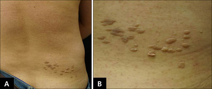
Fig. 1 (A) Multiple gray-brownish papules and confluent plaques with a zosteriform distribution on the right lower back. (B) Close-up view.
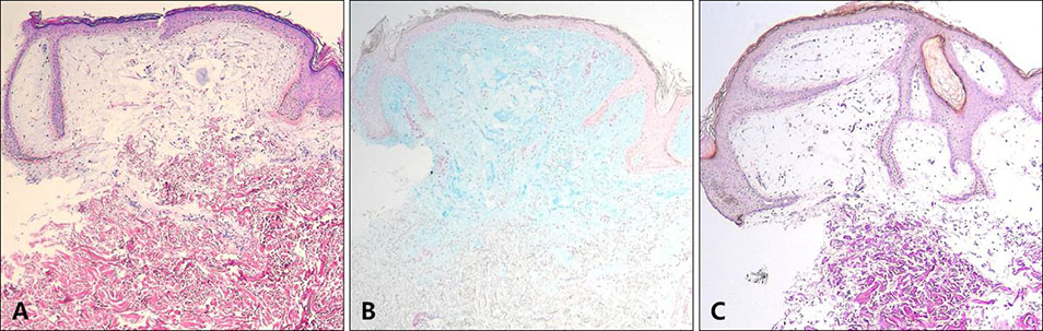
Fig. 2 (A) Amorphous materials and loosely separated collagen fibers in the papillary dermis and acanthosis with elongated rete ridges (H&E, ×40). (B) Positively stained bluish amorphous materials in the papillary dermis (Alcian blue at pH 2.5, ×40). (C) Reduced numbers of elastic fibers in the papillary dermis bearing mucin deposits (Verhoeff-van Gieson, ×40).
Reference
-
1. Redondo Bellón P, Vázquez-Doval J, Idoate M, Quintanilla E. Mucinous nevus. J Am Acad Dermatol. 1993; 28:797–798.
Article2. Rongioletti F, Rebora A. Mucinous nevus. Arch Dermatol. 1996; 132:1522–1523.
Article3. Cobos G, Braunstein I, Abuabara K, Chu EY, James W. Mucinous nevus: report of a case and review of the literature. JAMA Dermatol. 2014; 150:1018–1019.4. Tardío JC, Granados R. The cellular component of the mucinous nevus consists of CD34-positive fibroblasts. J Cutan Pathol. 2010; 37:1019–1020.
Article5. Chi CC, Wang SH, Lin PY. Combined epidermal-connective tissue nevus of proteoglycan (a type of mucinous nevus): a case report and literature review. J Cutan Pathol. 2009; 36:808–811.
Article6. Perez-Crespo M, Lopez-Navarro N, Betlloch I, Herrera E, Niveiro M, Gallego E. Acquired and familial mucinous nevus. Int J Dermatol. 2011; 50:1283–1285.
Article7. Vukicevic JS, Milobratovic DJ, Milinkovic MV, Bogdanovic Z. Extensive, adulthood inflammatory linear verrucous epidermal nevus associated with mucinous nevus. Indian J Dermatol Venereol Leprol. 2011; 77:607–608.
Article8. Song BH, Park S, Park EJ, Kwon IH, Kim KH, Kim KJ. Mucinous nevus with fat: an unusual case report and literature review. Am J Dermatopathol. 2012; 34:e146–e148.9. Kim EJ, Jo SJ, Cho KH. A case of mucinous nevus clinically mimicking nevus lipomatosus superficialis. Ann Dermatol. 2014; 26:549–550.
Article10. Sasaki T, Yoneda K, Yokoi I, Moriue J, Demitsu T, Kubota Y. Comorbidity of dermatofibromas and mucinous nevi. Int J Dermatol. 2016; 55:e53–e55.
Article11. Walter Lepage A, Frouin É, Junca A, Cante V, Monégier du Sorbier C, Hulin-Desquiret MC, et al. [Mucinous nevus of late onset]. Ann Dermatol Venereol. 2016; 143:547–553. French.12. Joo HJ, Yoo HJ, Kim JE, Kang H. A case of congenital mucinous nevus on the back. Korean J Dermatol. 2014; 52:892–894.13. Chen CW, Tsai TF, Chen YF, Hung CM. Familial mucinous nevus. Pediatr Dermatol. 2008; 25:288–289.
Article14. Lim JH, Cho SH, Kim HO, Kim CW, Park YM. Mucinous naevus with atypical features. Br J Dermatol. 2003; 148:1064–1066.
Article15. Brakman M, Starink TM, Tafelkruyer J, Bos JD. Linear connective tissue naevus of the proteoglycan type (‘naevus mucinosus’). Br J Dermatol. 1994; 131:368–370.
Article16. Yokogawa M, Kamakura T, Ishiguro H, Ikeda M, Kodama H. Mucinous nevus. J Dermatol. 2005; 32:30–33.
Article
- Full Text Links
-
- Actions
-
Cited
- CITED
-
- Close
- Share
-
- Similar articles
-
- A Case of Mucinous Nevus on Left Buttock Near the Anus
- A Case of Congenital Mucinous Nevus on the Back
- A Case of Mucinous Nevus Clinically Mimicking Nevus Lipomatosus Superficialis
- A Case of Acquired Mucinous Nevus in Nevus Lipomatosus Cutaneous Superficialis
- A Case of Bilateral Nevus of Ota Associated with Bilateral Nevus of Ito
