Ann Dermatol.
2014 Aug;26(4):549-550. 10.5021/ad.2014.26.4.549.
A Case of Mucinous Nevus Clinically Mimicking Nevus Lipomatosus Superficialis
- Affiliations
-
- 1Department of Dermatology, Seoul National University College of Medicine, Seoul, Korea. khcho@snu.ac.kr
- KMID: 2265611
- DOI: http://doi.org/10.5021/ad.2014.26.4.549
Abstract
- No abstract available.
Figure
-
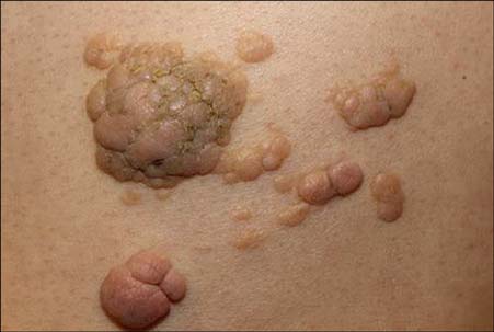
Fig. 1 Confluent flesh-colored to brownish non-firm papules and nodules on the left lower back, with a zosteriform distribution.
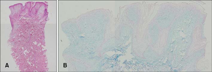
Fig. 2 (A) The band-like deposition of mucin was mainly observed in the superficial dermis, with papillomatosis, hyperkeratosis, and elongation of the rete ridge in the epidermis (H&E, ×40). (B) Mucin deposited in the papillary dermis stained positive to alcian blue at pH 2.5 (×100).
Cited by 1 articles
-
Mucinous Nevus
Min Young Lee, Ji Yeon Byun, Hae Young Choi, You Won Choi
Ann Dermatol. 2018;30(4):465-467. doi: 10.5021/ad.2018.30.4.465.
Reference
-
1. Chen CW, Tsai TF, Chen YF, Hung CM. Familial mucinous nevus. Pediatr Dermatol. 2008; 25:288–289.
Article2. Perez-Crespo M, Lopez-Navarro N, Betlloch I, Herrera E, Niveiro M, Gallego E. Acquired and familial mucinous nevus. Int J Dermatol. 2011; 50:1283–1285.
Article3. Song BH, Park S, Park EJ, Kwon IH, Kim KH, Kim KJ. Mucinous nevus with fat: an unusual case report and literature review. Am J Dermatopathol. 2012; 34:e146–e148.4. Chi CC, Wang SH, Lin PY. Combined epidermal-connective tissue nevus of proteoglycan (a type of mucinous nevus): a case report and literature review. J Cutan Pathol. 2009; 36:808–811.
Article
