Prediction and Staging of Hepatic Fibrosis in Children with Hepatitis C Virus: A Machine Learning Approach
- Affiliations
-
- 1Faculty of Informatics and Computer Science, The British University in Egypt, Cairo, Egypt. nahla.barakat@bue.edu.eg
- 2Department of Pediatrics, Faculty of Medicine, Alexandria University, Alexandria, Egypt.
- 3Ministry of Health, Alexandria, Egypt.
- KMID: 2457469
- DOI: http://doi.org/10.4258/hir.2019.25.3.173
Abstract
OBJECTIVES
The aim of this study is to develop an intelligent diagnostic system utilizing machine learning for data cleansing, then build an intelligent model and obtain new cutoff values for APRI (aspartate aminotransferase-to-platelet ratio) and FIB-4 (fibrosis score) for the prediction and staging of fibrosis in children with chronic hepatitis C (CHC).
METHODS
Random forest (RF) was utilized in this study for data cleansing; then, prediction and staging of fibrosis, APRI and FIB-4 scores and their areas under the ROC curve (AUC) have been obtained on the cleaned dataset. A cohort of 166 Egyptian children with CHC was studied.
RESULTS
RF, APRI, and FIB-4 achieved high AUCs; where APRI had AUCs of 0.78, 0.816, and 0.77; FIB-4 had AUCs of 0.74, 0.828, and 0.78; and RF had AUCs of 0.903, 0.894, and 0.822, for the prediction of any type of fibrosis, advanced fibrosis, and differentiating between mild and advanced fibrosis, respectively.
CONCLUSIONS
Machine learning is a valuable addition to non-invasive methods of liver fibrosis prediction and staging in pediatrics. Furthermore, the obtained cutoff values for APRI and FIB-4 showed good performance and are consistent with some previously obtained cutoff values. There was some agreement between the predictions of RF, APRI and FIB-4 for the prediction and staging of fibrosis.
MeSH Terms
Figure
-
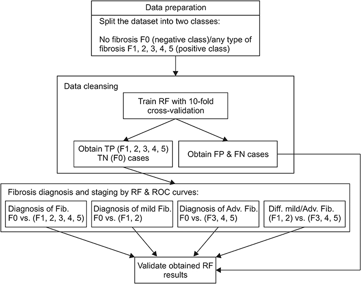
Figure 1 Steps in the proposed method. RF: random forest, TP: true positive, TN: true negative, FP: false positive, FN: false negative.
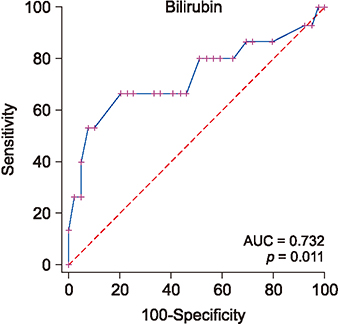
Figure 2 Bilirubin performance as a predictor of fibrosis.
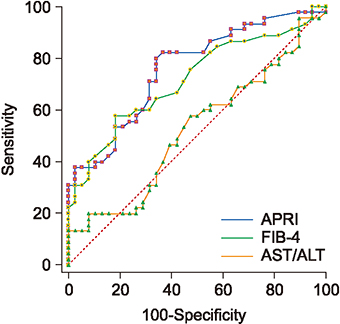
Figure 3 ROC curves for APRI, FIB-4, and AST/ALT ratio for predicting existence of any type of fibrosis. AST/ALT: alanine aminotransferase/aspartate aminotransferase, APRI: AST-to-platelet ratio, FIB-4: fibrosis score.
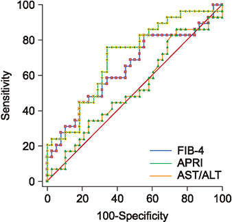
Figure 4 ROC curves for APRI, FIB-4, and AST/ALT ratio for predicting mild fibrosis. APRI has the best AUC, followed by FIB-4. AST/ALT: alanine aminotransferase/aspartate aminotransferase, APRI: AST-to-platelet ratio, FIB-4: fibrosis score, AUC: area under the ROC curve.
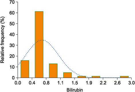
Figure 5 Bilirubin values distribution for no fibrosis/advanced fibrosis ≥0.9.
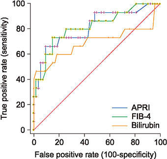
Figure 6 ROC curves for APRI, FIB-4 and bilirubin for predicting advanced fibrosis. FIB-4 has the best AUC followed by APRI. APRI: aspartate aminotransferase-to-platelet ratio, FIB-4: fibrosis score, AUC: area under the ROC curve.
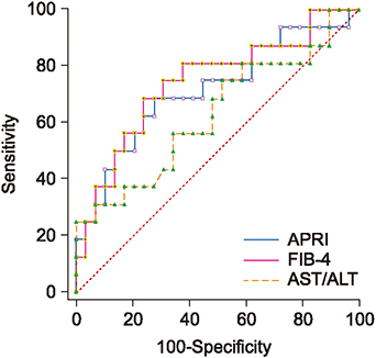
Figure 7 ROC curves for APRI, FIB-4, and AST/ALT for differentiating mild advanced fibrosis. FIB-4 has the best AUC followed by APRI. AST/ALT: alanine aminotransferase/aspartate aminotransferase, APRI: AST-to-platelet ratio, FIB-4: fibrosis score, AUC: area under the ROC curve.
Reference
-
1. World Health Organization. Global hepatitis report 2017. Geneva, Switzerland: World Health Organization;2017.2. Pokorska-Spiewak M, Kowalik-Mikołajewska B, Aniszewska M, Pluta M, Marczynska M. Is liver biopsy still needed in children with chronic viral hepatitis? World J Gastroenterol. 2015; 21(42):12141–12149.
Article3. Temple JL, Cordero P, Li J, Nguyen V, Oben JA. A guide to non-alcoholic fatty liver disease in childhood and adolescence. Int J Mol Sci. 2016; 17(6):E947.
Article4. Mansoor S, Collyer E, Alkhouri N. A comprehensive review of noninvasive liver fibrosis tests in pediatric nonalcoholic fatty liver disease. Curr Gastroenterol Rep. 2015; 17(6):23.
Article5. Valva P, Rios DA, De Matteo E, Preciado MV. Chronic hepatitis C virus infection: serum biomarkers in predicting liver damage. World J Gastroenterol. 2016; 22(4):1367–1381.
Article6. Stasi C, Milani S. Non-invasive assessment of liver fibrosis: between prediction/prevention of outcomes and cost-effectiveness. World J Gastroenterol. 2016; 22(4):1711–1720.
Article7. Kapogiannis BG, Leister E, Siberry GK, Van Dyke RB, Rudy B, Flynn P, et al. Prevalence of and progression to abnormal noninvasive markers of liver disease (aspartate aminotransferase-to-platelet ratio index and Fibrosis-4) among US HIV-infected youth. AIDS. 2016; 30(6):889–898.
Article8. Alkhouri N. Putting it all together: noninvasive diagnosis of fibrosis in nonalcoholic fatty liver disease in adults and children. Clin Liver Dis (Hoboken). 2017; 9(6):134–137.
Article9. Chen Y, Luo Y, Huang W, Hu D, Zheng RQ, Cong SZ, et al. Machine-learning-based classification of real-time tissue elastography for hepatic fibrosis in patients with chronic hepatitis B. Comput Biol Med. 2017; 89:18–23.
Article10. Brenner DA. Reversibility of liver fibrosis. Gastroenterol Hepatol (N Y). 2013; 9(11):737–739.11. Breiman L. Random forests. Mach Learn. 2001; 45(1):5–32.12. Elrazek A, Amer M, El-Hawary B, Salah A, Bhagavathula AS, Alboraie M, et al. Prediction of HCV vertical transmission: what factors should be optimized using data mining computational analysis. Liver Int. 2017; 37(4):529–533.
Article13. Hashem S, Esmat G, Elakel W, Habashy S, Raouf SA, Elhefnawi M, et al. Comparison of machine learning approaches for prediction of advanced liver fibrosis in chronic hepatitis C patients. IEEE/ACM Trans Comput Biol Bioinform. 2018; 15(3):861–868.
Article14. Awad A, Mabrouk M, Elakel W, Doss W, Awad T, Kamal S. Statistical and data mining analysis to identify clinical biochemical and pathological features of liver fibrosis versus Metavir score in a cohort of 69,106 chronic, hepatitis C patients in Egypt. Open Forum Infect Dis. 2016; 3:Suppl_1. 432.
Article15. El Raziky M, Awad AB, Youssef M, Awad A, Elshakawi A, Esmat G, et al. A novel prediction model for liver fibrosis in patients with chronic hepatitis c virus using Fibroscan and routine laboratory data. J Hepatol. 2013; 58:Suppl 1. S184.16. Barakat SH, El-Gandy W, Salem M, Ahmed N. Diagnostic algorithm for implementation of non-invasive scores for liver fibrosis in clinical practice in children with chronic hepatitis C. J Hepatol. 2015; 62:Suppl 2. S507.17. Barakat S, El-Gendy W, El-Naga HA, Salem M. Validation and comparison of six non-invasive scores for the diagnosis of liver fibrosis in children with chronic hepatitis C. J Hepatol. 2014; 60:1_Suppl. S312.18. Ishak K, Baptista A, Bianchi L, Callea F, De Groote J, Gudat F, et al. Histological grading and staging of chronic hepatitis. J Hepatol. 1995; 22(6):696–699.
Article19. Barakat NH, Bradley AP, Barakat MN. Intelligible support vector machines for diagnosis of diabetes mellitus. IEEE Trans Inf Technol Biomed. 2010; 14(4):1114–1120.
Article20. Ghaffar TA, Youssef A, Zalata K, ElSharkawy A, Mowafy M, Wanis AA, et al. Noninvasive assessment of liver fibrosis in Egyptian children with chronic liver diseases. Curr Pediat Res. 2016; 20(1-2):57–63.21. Poynard T, Ratziu V, Benmanov Y, Di Martino V, Bedossa P, Opolon P. Fibrosis in patients with chronic hepatitis C: detection and significance. Semin Liver Dis. 2000; 20(1):47–55.
Article22. Jackson JA, Konomi JV, Mendoza MV, Krasinskas A, Jin R, Caltharp S, et al. Performance of fibrosis prediction scores in paediatric non-alcoholic fatty liver disease. J Paediatr Child Health. 2018; 54(2):172–176.
Article23. Yang HR, Kim HR, Kim MJ, Ko JS, Seo JK. Noninvasive parameters and hepatic fibrosis scores in children with nonalcoholic fatty liver disease. World J Gastroenterol. 2012; 18(13):1525–1530.
Article24. Schmid P, Bregenzer A, Huber M, Rauch A, Jochum W, Mullhaupt B, et al. Progression of liver fibrosis in HIV/HCV co-infection: a comparison between non-invasive assessment methods and liver biopsy. PLoS One. 2015; 10(9):e0138838.
Article
- Full Text Links
-
- Actions
-
Cited
- CITED
-
- Close
- Share
-
- Similar articles
-
- Machine Learning vs. Statistical Model for Prediction Modelling: Application in Medical Imaging Research
- Prevention of Viral Hepatitis and Vaccination
- Toward hepatitis C virus elimination using artificial intelligence
- A Case of Coinfection of Hepatitis A and E Virus with Hepatic Encephalopathy
- Diagnostic Accuracy of Machine Learning Algorithms for Hepatitis A Antibody
