Computed Tomographic Features of Lung Parenchyma Over Time after Cardiopulmonary Resuscitation
- Affiliations
-
- 1Department of Radiology, Chungbuk National University Hospital, Cheongju, Korea. immdjy@gmail.com
- KMID: 2457451
- DOI: http://doi.org/10.3348/jksr.2019.80.4.740
Abstract
- PURPOSE
To identify the key CT features of lung parenchyma over time after cardiopulmonary resuscitation (CPR).
MATERIALS AND METHODS
In total, 72 patients underwent CT after CPR. Because the median time from return of spontaneous circulation (ROSC) to CT was 1 h 3 min, we divided patients into two groups: ≤ 1 h (group 1) and > 1 h (group 2), based on the ROSC to CT time. We analyzed and compared various lung parenchymal CT findings between groups.
RESULTS
Each group included 36 patients. Using statistical analysis, we identified seven statistically significant imaging features. Gradient (p = 0.010), lobular gradient (p = 0.017), diffuse pattern (p = 0.000), upper distribution (p = 0.032), and peripheral portion sparing (p = 0.000) were more common in group 1 than in group 2. Dependent density (p = 0.010) and lobular consolidation (p = 0.010) were more common in group 2 than in group 1.
CONCLUSION
The gradient and lobular gradient tended to disappear over time after ROSC. In terms of distribution, a diffuse pattern with upper predominance and peripheral portion sparing tended to disappear over time. However, the dependent density and lobular consolidation tended to increase over time in the lung parenchyma after CPR.
MeSH Terms
Figure
-

Fig. 1 CT features of a 91-year-old woman whose return of spontaneous circulation to CT interval was 17 min; lobular gradient in both upper lobes. On chest CT with lung setting, ground-glass attenuation is seen; this is nearly absent in the anterior part of the secondary pulmonary lobules and becomes thicker toward the dependent portion of the secondary pulmonary lobules (lobular gradient, arrows) in both upper lobes.
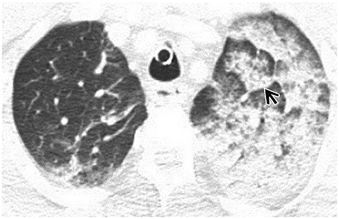
Fig. 2 CT features of a 53-year-old man whose return of spontaneous circulation to CT interval was 38 min; lobular gradient (arrow) and a gradient in the left upper lobe.
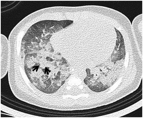
Fig. 3 CT features of a 1-year-old boy whose return of spontaneous circulation to CT interval was 1 h 22 min; lobular consolidation in the right middle and both lower lobes. On chest CT with lung setting, consolidation fills the secondary pulmonary lobules, without a gradient (arrows).

Fig. 4 CT features of a 79-year-old woman whose return of spontaneous circulation to CT interval was 2 h 21 min, with only dependent density in both lower lobes. On chest CT with lung settings, there are consolidations in dependent portion of both lower lobes (arrows).
Reference
-
1. Cho SH, Kim EY, Choi SJ, Kim YK, Sung YM, Choi HY, et al. Multidetector CT and radiographic findings of lung injuries secondary to cardiopulmonary resuscitation. Injury. 2013; 44:1204–1207.
Article2. Cha KC, Kim YW, Kim HI, Kim OH, Cha YS, Kim H, et al. Parenchymal lung injuries related to standard cardiopulmonary resuscitation. Am J Emerg Med. 2017; 35:117–121.
Article3. Wicky S, Wintermark M, Schnyder P, Capasso P, Denys A. Imaging of blunt chest trauma. Eur Radiol. 2000; 10:1524–1538.
Article4. Basu S, Nozari A, Liu XL, Rubertsson S, Wiklund L. Development of a novel biomarker of free radical damage in reperfusion injury after cardiac arrest. FEBS Lett. 2000; 470:1–6.
Article5. Muller NL. Blunt thoracic trauma. In : Muller NL, Silva CIS, editors. Imaging of the chest: expert radiology series. Philadelphia: Elsevier;2008. p. 1248–1249.6. Lomoschitz FM, Eisenhuber E, Linnau KF, Peloschek P, Schoder M, Bankier AA. Imaging of chest trauma: radiological patterns of injury and diagnostic algorithms. Eur J Radiol. 2003; 48:61–70.
Article
- Full Text Links
-
- Actions
-
Cited
- CITED
-
- Close
- Share
-
- Similar articles
-
- Liver Laceration with Hemoperitoneum after Cardiopulmonary Resuscitation
- Clinical Significance of Low-flow Time in Patients Treated with Extracorporeal Cardiopulmonary Resuscitation
- The Author's Response: Compression Rate during Cardiopulmonary Resuscitation
- Injuries by Resuscitation
- Cardiac Arrest Due to Unrecognized Congenital Diaphragmatic Hernia
