Hemarthrosis Occurred after Arthroscopic Rotator Cuff Repair in a Chronic Renal Failure Patient with a Stenosis in an Ipsilateral Arteriovenous Fistula
- Affiliations
-
- 1Department of Orthopaedic Surgery, St. Carollo Hospital, Suncheon, Korea. abeli@naver.com
- KMID: 2455506
- DOI: http://doi.org/10.4055/jkoa.2019.54.4.366
Abstract
- Hemarthrosis occurring after arthroscopic surgery for lesions of the shoulder joint is a very rare complication that can develop due to an injury to the blood vessels when an anterior portal is formed. This is a complication that rarely develops in patients who are taking antithrombotic drugs or who do not have associated diseases, such as thrombocytopenia. We report a case of hemarthrosis that occurred after performing arthroscopic surgery to repair a rotator cuff tear in a patient with a stenosis in an arteriovenous fistula for hemodialysis in the ipsilateral upper arm.
Keyword
MeSH Terms
Figure
-
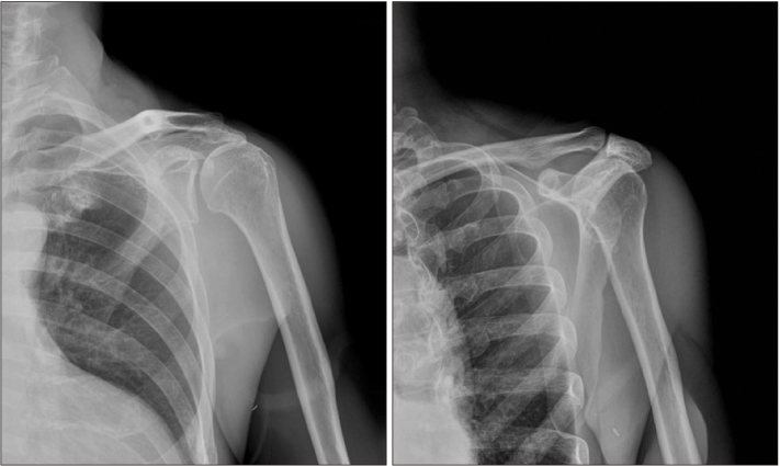
Figure 1 Plain radiographs showing the subacromial spur, proximal humeral migration and reduced acromiohumeral interval.
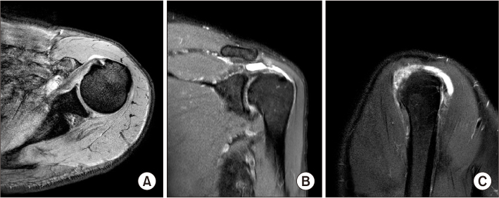
Figure 2 T2-weighted fat suppression magnetic resonance imaging. Axial (A), sagittal (B), and coronal (C) images showing a massive cuff tear with retraction.

Figure 3 Arthroscopic images. A partial subscapularis tear was observed and repaired using a single row technique (A), and full thickness tears of the supraspinatus and infraspinatus tendons were repaired using a suture bridge technique (B). There was no evidence of intra-articular bleeding during the procedure.

Figure 4 (A, B) On postoperative day 3, swelling was observed around the anterior portal, so joint aspiration was performed, and approximately 65 ml of dark-bloody fluid was drained. (C, D) Computed tomography angiography images. No dye leakage was observed, which implies vascular injury or a pseudoaneurysm, but a large collection of fluid around the joint was noted. Total occlusion of the cephalic vein (arrowhead) was observed, and subtotal stenosis of the axillary vein (empty arrow) and the outflow of the arteriovenous fistula (white arrow) was observed.

Figure 5 (A) Gross photograph of the arteriovenous fistula on the upper arm before ligation of the arteriovenous (AV) fistula. (B) After ligation of the AV fistula, a permanent catheter for dialysis was inserted through the contralateral internal jugular vein.
Reference
-
1. Lo IK, Lind CC, Burkhart SS. Glenohumeral arthroscopy portals established using an outside-in technique: neurovascular anatomy at risk. Arthroscopy. 2004; 20:596–602.
Article2. Curtis AS, Snyder SJ, Del Pizzo W, Friedman MJ, Ferkel RD, Karzel RP. Complications of shoulder arthroscopy. Arthroscopy. 1992; 8:395.3. Cameron SE. Venous pseudoaneurysm as a complication of shoulder arthroscopy. J Shoulder Elbow Surg. 1996; 5:404–406.
Article4. Godin JA, Mayer SW, Garrigues GE, Mather RC 3rd. Pseudoaneurysm after shoulder arthroscopy. J Shoulder Elbow Surg. 2013; 22:e12–e17.
Article5. Hall HC, Moudgill N, Kahn M, et al. An unusual cause of venous hypertension after dialysis access creation. Ann Vasc Surg. 2011; 25:983.e1–983.e4.
Article6. Neville RF, Abularrage CJ, White PW, Sidawy AN. Venous hypertension associated with arteriovenous hemodialysis access. Semin Vasc Surg. 2004; 17:50–56.
Article7. Small NC. Complications in arthroscopic surgery performed by experienced arthroscopists. Arthroscopy. 1988; 4:215–221.
Article8. Tsujii A, Tanaka Y, Yonetani Y, Shiozaki Y, Tomiyama Y, Horibe S. Knee hemarthrosis after arthroscopic surgery in an athlete with low factor XIII activity. Sports Med Arthrosc Rehabil Ther Technol. 2012; 4:35.
Article9. Im JH, Huh SW, Park MK, Lee JY. Volar loking plate fixation for distal radius fractures in hemodialysis patients. J Korean Soc Surg Hand. 2015; 20:96–103.
- Full Text Links
-
- Actions
-
Cited
- CITED
-
- Close
- Share
-
- Similar articles
-
- Revisional Rotator Cuff Repair
- New Retear Pattern after Rotator Cuff Repair at Previous Intact Portion of Rotator Cuff
- Surgical Options for Failed Rotator Cuff Repair, except Arthroplasty: Review of Current Methods
- Various Regimens for the Functional Recovery after Arthroscopic Shoulder Surgery
- Arthroscopic Bony Procedure During of Rotator Cuff Repair: Acromioplasty, Distal Clavicle Resection, Footprint Preparation and Coracoplasty
