Korean J Transplant.
2019 Jun;33(2):30-35. 10.4285/jkstn.2019.33.2.30.
Diagnostic performance of multidetector computed tomography for symptomatic lymphoceles in kidney transplant recipients
- Affiliations
-
- 1Department of Surgery, Ilsan Paik Hospital, Inje University College of Medicine, Goyang, Korea. midasia@hanmail.net
- 2Department of Radiology, Korea University Anam Hospital, Korea University College of Medicine, Seoul, Korea.
- KMID: 2453796
- DOI: http://doi.org/10.4285/jkstn.2019.33.2.30
Abstract
- BACKGROUND
To evaluate the size of a postoperative lymphocele in the coronal and axial reconstruction planes using multidetector computed tomography (MDCT) in kidney transplantation recipients.
METHODS
We evaluated 92 recipients who underwent MDCT of the abdominopelvis at 1 month after kidney transplantation. The axial short axis, axial surface area, coronal short axis, and coronal surface area of the lymphocele were measured using the reconstructed MDCT coronal and axial images. Depending on the clinical manifestations and radiologic findings of the recipients, all lymphoceles were classified into symptomatic and asymptomatic. We compared the suitability of the size measurement on coronal and axial planes of MDCT reconstruction for symptomatic lymphocele in kidney transplant recipients using Spearman's correlation analysis and comparisons of receiver operating characteristic (ROC) curves.
RESULTS
Areas under the ROC curves were 0.957 and 0.928 for the axial short axis and axial surface area and 0.968 and 0.966 for the coronal short axis and coronal surface area, respectively. In pairwise comparison of the ROC curve of the parameters of the symptomatic lymphoceles, the coronal measurement was significant in contrast to the axial measurement (short axis, P=0.357; surface area, P=0.047).
CONCLUSIONS
For the prediction of symptomatic lymphoceles using MDCT, the coronal measurement of postoperative lymphoceles can significantly improve diagnostic performance over axial measurement in kidney transplant recipients.
MeSH Terms
Figure
-
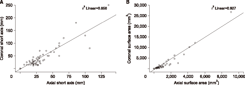
Fig. 1 Linear regression analysis of lymphocele parameters between the axial and coronal planes. (A) The correlation between the axial and coronal short axes had a Pearson's coefficient of 0.925. (B) The correlation between the axial and coronal surface areas had a Pearson's coefficient of 0.962.
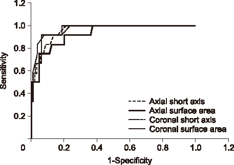
Fig. 2 Receiver operating characteristic curve and cutoff value for a symptomatic lymphocele that requires radiologic or surgical intervention.
Reference
-
1. Hariharan S, Johnson CP, Bresnahan BA, Taranto SE, McIntosh MJ, Stablein D. Improved graft survival after renal transplantation in the United States, 1988 to 1996. N Engl J Med. 2000; 342:605–612.
Article2. Zietek Z, Sulikowski T, Tejchman K, Sieńko J, Janeczek M, Iwan-Zietek I, et al. Lymphocele after kidney transplantation. Transplant Proc. 2007; 39:2744–2747.
Article3. Ebadzadeh MR, Tavakkoli M. Lymphocele after kidney transplantation: where are we standing now? Urol J. 2008; 5:144–148.4. Ulrich F, Niedzwiecki S, Fikatas P, Nebrig M, Schmidt SC, Kohler S, et al. Symptomatic lymphoceles after kidney transplantation: multivariate analysis of risk factors and outcome after laparoscopic fenestration. Clin Transplant. 2010; 24:273–280.
Article5. Giuliani S, Gamba P, Kiblawi R, Midrio P, Ghirardo G, Zanon GF. Lymphocele after pediatric kidney transplantation: incidence and risk factors. Pediatr Transplant. 2014; 18:720–725.
Article6. Gomes AS, Scholl D, Feinberg S, Simmons RL, Amplatz K. Lymphangiography and ultrasound in management of lymphoceles. Urology. 1979; 13:104–108.
Article7. Goel M, Flechner SM, Zhou L, Mastroianni B, Savas K, Derweesh I, et al. The influence of various maintenance immunosuppressive drugs on lymphocele formation and treatment after kidney transplantation. J Urol. 2004; 171:1788–1792.
Article8. Chedid MF, Muthu C, Nyberg SL, Lesnick TG, Kremers WK, Prieto M, et al. Living donor kidney transplantation using laparoscopically procured multiple renal artery kidneys and right kidneys. J Am Coll Surg. 2013; 217:144–152.
Article9. Adani GL, Baccarani U, Bresadola V, Lorenzin D, Montanaro D, Risaliti A, et al. Graft loss due to percutaneous sclerotherapy of a lymphocele using acetic acid after renal transplantation. Cardiovasc Intervent Radiol. 2005; 28:836–838.
Article10. Król R, Kolonko A, Chudek J, Ziaja J, Pawlicki J, Mały A, et al. Did volume of lymphocele after kidney transplantation determine the choice of treatment modality? Transplant Proc. 2007; 39:2740–2743.
Article11. Sim A, Ng LG, Cheng C. Occurrence of a lymphocele following renal transplantation. Singapore Med J. 2013; 54:259–262.
Article12. Yu YL, Lee MS, Juan CJ, Hueng DY. Calculating the tumor volume of acoustic neuromas: comparison of ABC/2 formula with planimetry method. Clin Neurol Neurosurg. 2013; 115:1371–1374.
Article13. Saura D, de la Morena G, Flores-Blanco PJ, Oliva MJ, Caballero L, González-Carrillo J, et al. Aortic valve stenosis planimetry by means of three-dimensional transesophageal echocardiography in the real clinical setting: feasibility, reliability and systematic deviations. Echocardiography. 2015; 32:508–515.
Article14. Dowlatshahi D, Kosior JC, Idris S, Eesa M, Dickhoff P, Joshi M, et al. Planimetric hematoma measurement in patients with intraventricular hemorrhage: is total volume a preferred target for reliable analysis? Stroke. 2012; 43:1961–1963.
Article15. Guinet C, Rousset P, Bobbio A, Alifano M, Damotte D, Régnard JF, et al. Comparison of coronal and axial computed tomography measurements of mediastinal nodes before primary surgery for non-small cell lung cancer. Eur J Radiol. 2012; 81:2440–2443.
Article
- Full Text Links
-
- Actions
-
Cited
- CITED
-
- Close
- Share
-
- Similar articles
-
- Laparoscopic Intraperitoneal Drainage of Lymphocele Developed Adjacent to the Kidney Transplanted
- Successful treatment of symptomatic lymphocele after kidney transplantation
- Evaluation of postoperative lymphocele according to amounts and symptoms by using 3-dimensional CT volumetry in kidney transplant recipients
- A Concept Analysis of Compliance in Kidney Transplant Recipient Including Compliance with Immunosuppressive Medication
- Correlation of allograft size versus body mass index to the incidence of hyperfiltration injury among kidney transplant recipients
