Comparison of tooth movement and biological response in corticotomy and micro-osteoperforation in rabbits
- Affiliations
-
- 1Graduate School, The Catholic University of Korea, Seoul, Korea.
- 2Department of Orthodontics, Seoul St. Mary's Hospital, College of Medicine, The Catholic University of Korea, Seoul, Korea. kook2002@catholic.ac.kr
- 3Department of Preventive Dentistry, College of Dentistry, King Faisal University, Al-hofuf, Saudi Arabia.
- 4Postgraduate Orthodontic Program, Arizona School of Dentistry & Oral Health, A.T. Still University, Mesa, AZ, USA.
- 5Graduate School of Dentistry, Kyung Hee University, Seoul, Korea.
- 6Department of Dentistry, Uijeongbu St. Mary's Hospital, College of Medicine, The Catholic University of Korea, Uijeongbu, Korea.
- 7Department of Orthodontics, Faculty of Dentistry, Ain Shams University, Cairo, Egypt.
- KMID: 2453057
- DOI: http://doi.org/10.4041/kjod.2019.49.4.205
Abstract
OBJECTIVE
The aim of this study was to evaluate the amount of tooth movement and histologic changes with different corticotomy designs and micro-osteoperforation in rabbits.
METHODS
The sample consisted of 24 rabbits divided into three experimental groups (triangular corticotomy [TC] and indentation corticotomy [IC] with flap, and flapless micro-osteoperforations [MP]) and a control. A traction force of 100 cN was applied by connecting the first premolars to the incisors. The amount of tooth movement was measured. Kruskal-Wallis test was used to assess differences in tooth movement between the groups. Micro-computed tomography, hematoxylin and eosin staining, and tartrate-resistant acidic phosphatase (TRAP) analysis were performed. Analysis of variance was applied to assess differences in TRAP-positive osteoclast count between the groups.
RESULTS
The amount of tooth movement increased by 46.5% and 32.0% in the IC and MP groups, respectively, while the bone fraction analysis showed 69.7% and 8.5% less mineralization compared to the control. There were no significant intergroup differences in the number of TRAP-positive osteoclasts.
CONCLUSIONS
The micro-osteoperforation group showed no significant differences in the amount of tooth movement compared to the corticotomy groups, nor in the TRAP-positive osteoclast count compared to both corticotomy groups and control.
Keyword
MeSH Terms
Figure
-
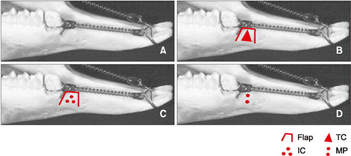
Figure 1 Different study groups. A, Control group (no surgical intervention); B, triangular corticotomy (TC) group; C, indentation corticotomy (IC) group; D, micro-osteoperforation (MP) group.
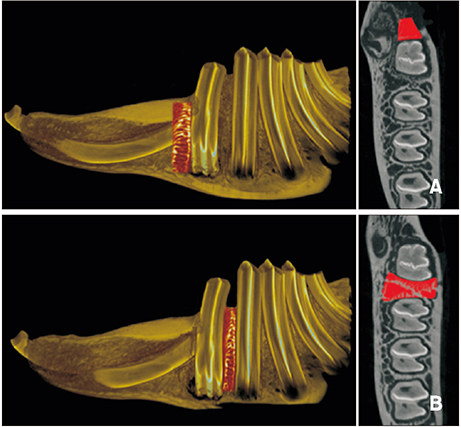
Figure 2 Specimen sites for micro-computed tomography imaging. A, The intervention site, mesial area of the first premolar; B, the non-intervention site, the area between the first and second premolars.
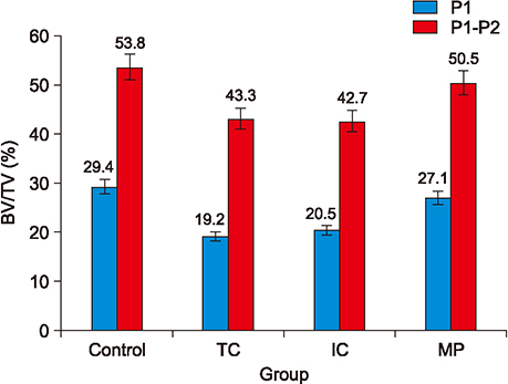
Figure 3 Percentage of bone volume (BV)/tissue volume (TV) in the four groups. P1, Mesial area of the first premolar; P1–P2, area between the first and second premolars; TC, triangular corticotomy; IC, indentation corticotomy; MP, micro-osteoperforation.

Figure 4 Microphotograph of periodontal tissues with tartrate-resistant acidic phosphatase (TRAP) staining (×200). A, Control group; B, triangular corticotomy (TC) group; C, indentation corticotomy group; D, micro-osteoperforation group. TRAP-positive osteoclasts can be observed on the compression side along the resorbed surface of the alveolar bone. The TC group shows several clusters of TRAP-positive osteoclasts, while the control group forms a straight band.
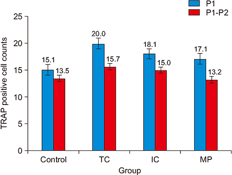
Figure 5 Comparison of tartrate-resistant acidic phosphatase (TRAP)-positive osteoclast counts. P1, Mesial area of the first premolar; P1–P2, area between the first and second premolars; TC, triangular corticotomy; IC, indentation corticotomy; MP, micro-osteoperforation.
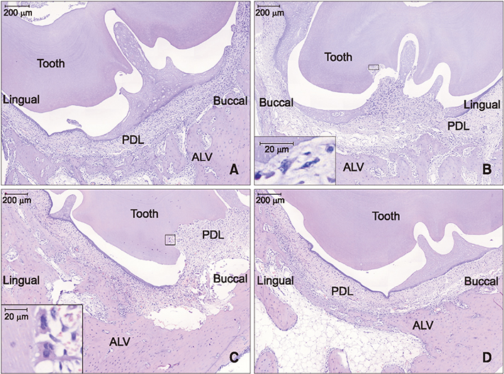
Figure 6 Microphotograph of buccolingual section of the mesial periodontium of the first premolar with H&E staining (×40) of decalcified specimens. A, Control group; B, triangular corticotomy (TC) group; C, indentation corticotomy (IC) group; D, micro-osteoperforation group. Note the enlarged periodontal ligament (PDL) space in B and C, and the increased number of odontoclasts on some teeth surfaces, especially in the TC (B) and IC (C) groups, which might have caused the enlarged PDL space by its increased activity. The figure suggests elevated root resorption, particularly in the TC and IC groups. ALV, Alveolar bone.
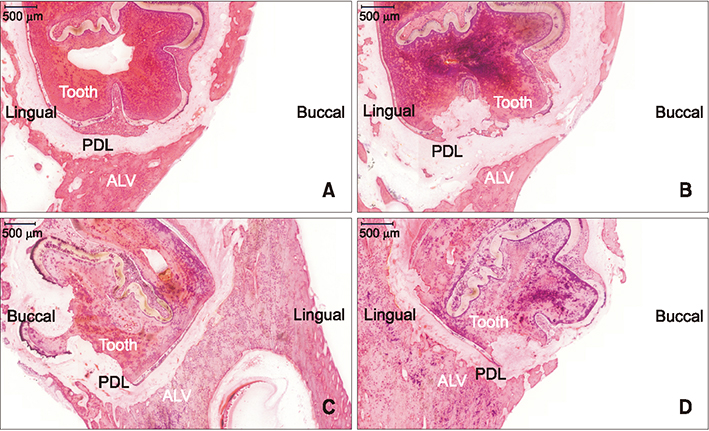
Figure 7 Microphotograph of buccolingual section of the mesial periodontium of the first premolar with H&E staining (×40) of undecalcified specimens. A, Control group; B, triangular corticotomy (TC) group; C, indentation corticotomy (IC) group; D, micro-osteoperforation (MP) group. There was more pronounced root and alveolar bone resorption in the TC (B) and the IC group (C) compared to the MP group (D). PDL, Periodontal ligament; ALV, alveolar bone.
Cited by 1 articles
-
Comparison of the effects of horizontal and vertical micro-osteoperforations on the biological response and tooth movement in rabbits
Seok-gon Kim, Yoon-Ah Kook, Hee Jin Lim, Patrick Park, Won Lee, Jae Hyun Park, Mohamed Bayome, Yoonji Kim
Korean J Orthod. 2021;51(5):304-312. doi: 10.4041/kjod.2021.51.5.304.
Reference
-
1. Igarashi K, Mitani H, Adachi H, Shinoda H. Anchorage and retentive effects of a bisphosphonate (AHBuBP) on tooth movements in rats. Am J Orthod Dentofacial Orthop. 1994; 106:279–289.
Article2. Işeri H, Kişnişci R, Bzizi N, Tüz H. Rapid canine retraction and orthodontic treatment with dentoalveolar distraction osteogenesis. Am J Orthod Dentofacial Orthop. 2005; 127:533–541. quiz 625.
Article3. Liou EJ, Huang CS. Rapid canine retraction through distraction of the periodontal ligament. Am J Orthod Dentofacial Orthop. 1998; 114:372–382.
Article4. Gantes B, Rathbun E, Anholm M. Effects on the periodontium following corticotomy-facilitated orthodontics. Case reports. J Periodontol. 1990; 61:234–238.
Article5. Chung KR, Oh MY, Ko SJ. Corticotomy-assisted orthodontics. J Clin Orthod. 2001; 35:331–339.6. Suya H. Corticotomy in orthodontics. In : Hosl E, Baldauf A, editors. Mechanical and biological basics in orthodontic therapy. Heidelberg: Huthig Buch Verlag;1991. p. 207–226.7. Kook YA, Lee W, Kim SH, Chung KR. Corticotomy-assisted space closure in adult patients with missing lower molars. J Clin Orthod. 2013; 47:85–95. quiz 139.8. McBride MD, Campbell PM, Opperman LA, Dechow PC, Buschang PH. How does the amount of surgical insult affect bone around moving teeth? Am J Orthod Dentofacial Orthop. 2014; 145:S92–S99.
Article9. Mostafa YA, Mohamed Salah, Mehanni S, ElBokle NN, Heider AM. Comparison of corticotomy-facilitated vs standard tooth-movement techniques in dogs with miniscrews as anchor units. Am J Orthod Dentofacial Orthop. 2009; 136:570–577.
Article10. Teixeira CC, Khoo E, Tran J, Chartres I, Liu Y, Thant LM, et al. Cytokine expression and accelerated tooth movement. J Dent Res. 2010; 89:1135–1141.
Article11. Alikhani M, Raptis M, Zoldan B, Sangsuwon C, Lee YB, Alyami B, et al. Effect of micro-osteoperforations on the rate of tooth movement. Am J Orthod Dentofacial Orthop. 2013; 144:639–648.
Article12. Cassetta M, Altieri F, Pandolfi S, Giansanti M. The combined use of computer-guided, minimally invasive, flapless corticotomy and clear aligners as a novel approach to moderate crowding: a case report. Korean J Orthod. 2017; 47:130–141.
Article13. Charavet C, Lecloux G, Bruwier A, Rompen E, Maes N, Limme M, et al. Localized piezoelectric alveolar decortication for orthodontic treatment in adults: a randomized controlled trial. J Dent Res. 2016; 95:1003–1009.
Article14. Ruso S, Campbell PM, Rossmann J, Opperman LA, Taylor RW, Buschang PH. Bone response to buccal tooth movements-with and without flapless alveolar decortication. Eur J Orthod. 2014; 36:613–623.
Article15. Swapp A, Campbell PM, Spears R, Buschang PH. Flapless cortical bone damage has no effect on medullary bone mesial to teeth being moved. Am J Orthod Dentofacial Orthop. 2015; 147:547–558.
Article16. Lindskog-Stokland B, Hansen K, Ekestubbe A, Wennström JL. Orthodontic tooth movement into edentulous ridge areas--a case series. Eur J Orthod. 2013; 35:277–285.
Article17. Nagaraj K, Upadhyay M, Yadav S. Titanium screw anchorage for protraction of mandibular second molars into first molar extraction sites. Am J Orthod Dentofacial Orthop. 2008; 134:583–591.
Article18. Santos PBDD, Herrera Sanches FS, Ferreira MC, de Almeida ALPF, Janson G, Garib D. Movement of mandibular molar into edentulous alveolar ridge: a cone-beam computed tomography study. Am J Orthod Dentofacial Orthop. 2017; 151:907–913.
Article19. Kim T, Handa A, Iida J, Yoshida S. RANKL expression in rat periodontal ligament subjected to a continuous orthodontic force. Arch Oral Biol. 2007; 52:244–250.
Article20. Yu JY, Lee W, Park JH, Bayome M, Kim Y, Kook YA. Histologic effects of intentional-socket-assisted orthodontic movement in rabbits. Korean J Orthod. 2012; 42:207–217.
Article21. Chen YW, Wang HC, Gao LH, Liu C, Jiang YX, Qu H, et al. Osteoclastogenesis in local alveolar bone in early decortication-facilitated orthodontic tooth movement. PLoS One. 2016; 11:e0153937.
Article22. Alkebsi A, Al-Maaitah E, Al-Shorman H, Abu Alhaija E. Three-dimensional assessment of the effect of micro-osteoperforations on the rate of tooth movement during canine retraction in adults with Class II malocclusion: a randomized controlled clinical trial. Am J Orthod Dentofacial Orthop. 2018; 153:771–785.
Article23. Cheung T, Park J, Lee D, Kim C, Olson J, Javadi S, et al. Ability of mini-implant-facilitated micro-osteoperforations to accelerate tooth movement in rats. Am J Orthod Dentofacial Orthop. 2016; 150:958–967.
Article24. Buschang PH. Surgically facilitated orthodontics: what does the evidence say?. In : Kapila SD, Goonewardene M, Kim-Berman H, Koster KY, editors. Interdisciplinary therapy: using contemporary approaches for complex cases. Ann Arbor: The University of Michigan;2016. p. 135–165.25. Aboul-Ela SM, El-Beialy AR, El-Sayed KM, Selim EM, El-Mangoury NH, Mostafa YA. Miniscrew implant-supported maxillary canine retraction with and without corticotomy-facilitated orthodontics. Am J Orthod Dentofacial Orthop. 2011; 139:252–259.
Article26. Roberts WE. Bone tissue interface. Int J Oral Implantol. 1988; 5:71–74.
Article27. Mapara M, Thomas BS, Bhat KM. Rabbit as an animal model for experimental research. Dent Res J (Isfahan). 2012; 9:111–118.
Article28. Castañeda S, Largo R, Calvo E, Rodríguez-Salvanés F, Marcos ME, Díaz-Curiel M, et al. Bone mineral measurements of subchondral and trabecular bone in healthy and osteoporotic rabbits. Skeletal Radiol. 2006; 35:34–41.
Article29. Chan E, Dalci O, Petocz P, Papadopoulou AK, Darendeliler MA. Physical properties of root cementum: part 26. Effects of micro-osteoperforations on orthodontic root resorption: a microcomputed tomography study. Am J Orthod Dentofacial Orthop. 2018; 153:204–213.
Article30. Owen KM, Campbell PM, Feng JQ, Dechow PC, Buschang PH. Elevation of a full-thickness mucoperiosteal flap alone accelerates orthodontic tooth movement. Am J Orthod Dentofacial Orthop. 2017; 152:49–57.
Article
- Full Text Links
-
- Actions
-
Cited
- CITED
-
- Close
- Share
-
- Similar articles
-
- Corticotomy and the Intrusive Tooth Movement
- Comparison of the effects of horizontal and vertical micro-osteoperforations on the biological response and tooth movement in rabbits
- Corticotomy and the molar uprighting
- Corticotomy for orthodontic tooth movement
- The effectiveness of corticotomy and piezocision on canine retraction: A systematic review
