Undifferentiated Pleomorphic Sarcoma of the Descending Thoracic Aorta Mimicking Pseudoaneurysm with Periaortic Hematoma: a Case Report
- Affiliations
-
- 1Department of Radiology, Hallym University Dongtan Sacred Heart Hospital, Hwaseong, Gyeonggi-do, Korea. yabae@hallym.or.kr
- 2Department of Pathology, Hallym University Dongtan Sacred Heart Hospital, Hwaseong, Gyeonggi-do, Korea.
- KMID: 2452531
- DOI: http://doi.org/10.13104/imri.2019.23.2.162
Abstract
- Undifferentiated pleomorphic sarcoma (UPS) arising from the descending thoracic aorta is a rare type of tumor. To our knowledge, only a few cases have been reported in the literature. We present computed tomography (CT) and magnetic resonance imaging findings of a 43-year-old male patient with undifferentiated pleomorphic sarcoma of the descending thoracic aorta, which showed enhancement on only magnetic resonance imaging (MRI). MRI with contrast enhancement may be useful in differentiating an aortic tumor from atherosclerotic disease.
MeSH Terms
Figure
-
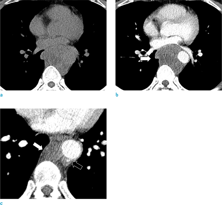
Fig. 1 Chest CT images of a 43-year-old man with undifferentiated pleomorphic sarcoma of the descending thoracic aorta. Axial pre- (a) and post-contrast enhanced (b, c) images show heterogeneous attenuated mass broadly attached to the descending thoracic aorta (white arrows). The mass shows no significant enhancement on CT images. There is a small ulcer-like projection at the inferior portion of the mass (black arrow on c).
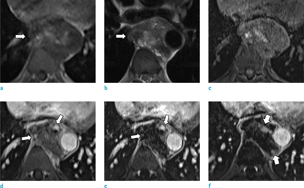
Fig. 2 Thoracic MR images of the patient. Axial T1-weighted (a) and T2-weighted (b) images show the mass of the descending thoracic aorta with heterogeneous signal intensity (white arrows). Axial T1-weighted, fat-saturated pre- (c) and post-contrast image (d), subtraction images (e, f) show peripheral rim and small nodular enhancement of the mass (white arrows), which was not shown on CT images.
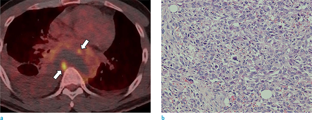
Fig. 3 PET-CT and microscopic findings of the patient. Axial PET-CT image (a) shows the FDG hypermetabolism on the periphery of the mass (white arrows), corresponding to enhancing portions on MRI. The pathologic finding (b) shows that the tumor is composed of short spindle pleomorphic cells with prominent nucleoli and high mitotic rates (Hematoxylin & Eosin stain, × 40).
Reference
-
1. Sebenik M, Ricci A Jr, DiPasquale B, et al. Undifferentiated intimal sarcoma of large systemic blood vessels: report of 14 cases with immunohistochemical profile and review of the literature. Am J Surg Pathol. 2005; 29:1184–1193.2. von Falck C, Meyer B, Fegbeutel C, et al. Imaging features of primary sarcomas of the great vessels in CT, MRI and PET/CT: a single-center experience. BMC Med Imaging. 2013; 13:25.
Article3. Heo SY, Park CS, Kim SJ, Park NH, Heo JH, Lee JJ. Undifferentiated pleomorphic sarcoma of the thoracic aorta: a case report. J Korean Soc Radiol. 2016; 75:304–308.
Article4. Goldblum JR. An approach to pleomorphic sarcomas: can we subclassify, and does it matter? Mod Pathol. 2014; 27:Suppl 1. S39–S46.
Article5. Chiche L, Mongredien B, Brocheriou I, Kieffer E. Primary tumors of the thoracoabdominal aorta: surgical treatment of 5 patients and review of the literature. Ann Vasc Surg. 2003; 17:354–364.
Article6. Wright EP, Glick AD, Virmani R, Page DL. Aortic intimal sarcoma with embolic metastases. Am J Surg Pathol. 1985; 9:890–897.
Article7. Thalheimer A, Fein M, Geissinger E, Franke S. Intimal angiosarcoma of the aorta: report of a case and review of the literature. J Vasc Surg. 2004; 40:548–553.
Article8. Utsunomiya D, Ikeda O, Ideta I, Hirayama T, Yamashita Y, Kamio T. Malignant fibrous histiocytoma arising from the aortic wall mimicking a pseudoaneurysm with ulceration. Circ J. 2007; 71:1659–1661.
Article9. Dedeilias P, Koletsis E, Nenekidis I, et al. Intimal aortic sarcoma mimicking ruptured thoracoabdominal type IV aneurysm. A rare case report and review of the literature. J Cardiothorac Surg. 2011; 6:162.
Article10. Rusthoven CG, Liu AK, Bui MM, et al. Sarcomas of the aorta: a systematic review and pooled analysis of published reports. Ann Vasc Surg. 2014; 28:515–525.
Article
- Full Text Links
-
- Actions
-
Cited
- CITED
-
- Close
- Share
-
- Similar articles
-
- Undifferentiated Pleomorphic Sarcoma of the Thoracic Aorta: A Case Report
- Undifferentiated Pleomorphic Sarcoma of the Thoracic Aorta Presenting with Ruptured Saccular Aneurysm: A Case Report
- A Case of Pleomorphic Dermal Sarcoma Showing Characteristics of Myxoinflammatory Fibroblastic Sarcoma
- Cutaneous Metastatic Undifferentiated Pleomorphic Sarcoma from a Mediastinal Sarcoma
- Intramural Hematoma of the Descending Thoracic Aorta Misdiagnosed as Aortic Rupture: A case report
