Comparison of different impression techniques for edentulous jaws using three-dimensional analysis
- Affiliations
-
- 1Department of Prosthodontics, School of Dentistry, Chonnam National University, Gwangju, Republic of Korea. psw320@naver.com
- KMID: 2450994
- DOI: http://doi.org/10.4047/jap.2019.11.3.179
Abstract
- PURPOSE
The purpose of this study was to compare two novel impression methods and a conventional impression method for edentulous jaws using 3-dimensional (3D) analysis software.
MATERIALS AND METHODS
Five edentulous patients (four men and one woman; mean age: 62.7 years) were included. Three impression techniques were used: conventional impression method (CI; control), simple modified closed-mouth impression method with a novel tray (SI), and digital impression method using an intraoral scanner (DI). Subsequently, a gypsum model was made, scanned, and superimposed using 3D analysis software. Mean area displacement was measured using CI method to evaluate differences in the impression surfaces as compared to those values obtained using SI and DI methods. The values were confirmed at two to five areas to determine the differences. CI and SI were compared at all areas, while CI and DI were compared at the supporting areas. Kruskal-Wallis test was performed for all data. Statistical significance was considered at P value <.05.
RESULTS
In the comparison of the CI and SI methods, the greatest difference was observed in the mandibular vestibule without statistical significance (P>.05); the difference was < 0.14 mm in the maxilla. The difference in the edentulous supporting areas between the CI and DI methods was not significant (P>.05).
CONCLUSION
The CI, SI, and DI methods were effective in making impressions of the supporting areas in edentulous patients. The SI method showed clinically applicability.
Keyword
Figure
-
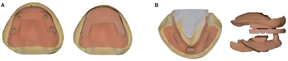
Fig. 1 (A) A novel maxillary tray for a simple modified impression method. (left) Not containing an occlusal rim; four-point support may be connected to the occlusal plate, but allowing slight movement of the plate to aid in locating the maxillary anterior teeth. (right) Occlusal plate with applied rim; unlike the conventional occlusal rim, this is designed to be easy to use with a large surface area to record the bite or for gothic arch tracing. (B) A novel mandibular tray for a simple modified impression method. (left) Occlusal view, (right) lateral view of both the novel maxillary and mandibular trays; the closed-mouth technique is performed by biting the maxillary occlusal plate and the mandibular anterior short occlusal rim using the bimanual method. The plate and mandibular occlusal rim have indentations so that the silicone bite registration material is precisely positioned between the two trays.
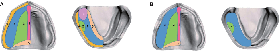
Fig. 2 (A) Areas compared by the CI and SI methods; (left) 1. Medial palatine raphe, 2. Hard palate, 3. Residual ridge, 4. Buccal vestibule, 5. Soft palate, (right) 1. Residual ridge, 2. Buccal shelf, 3. Retromolar pad, 4. Buccal vestibule, 5. Labial vestibule. (B) Areas compared by the CI and DI methods; (left) 1. Medial palatine raphe, 2. Hard palate, 3. Residual ridge, 4. Soft palate, (right) 1. Residual ridge, 2. Buccal shelf.
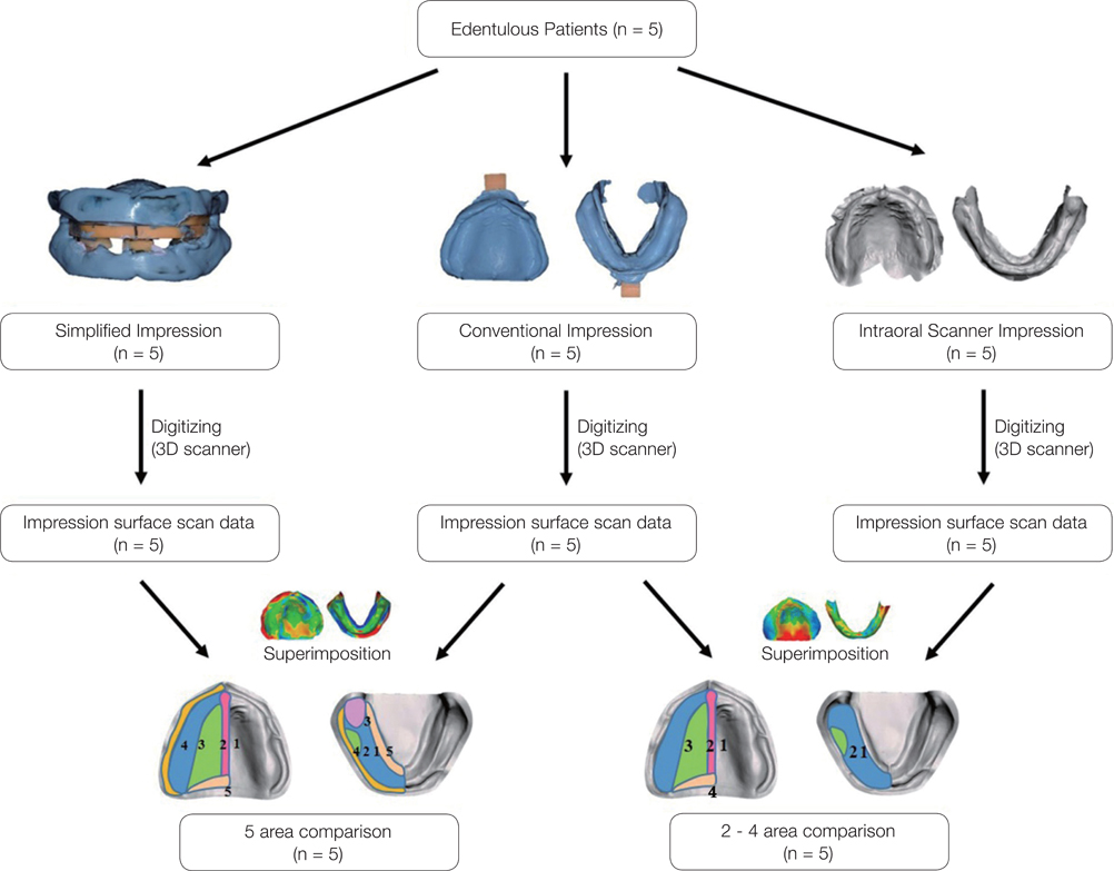
Fig. 3 Protocol of this experiment.
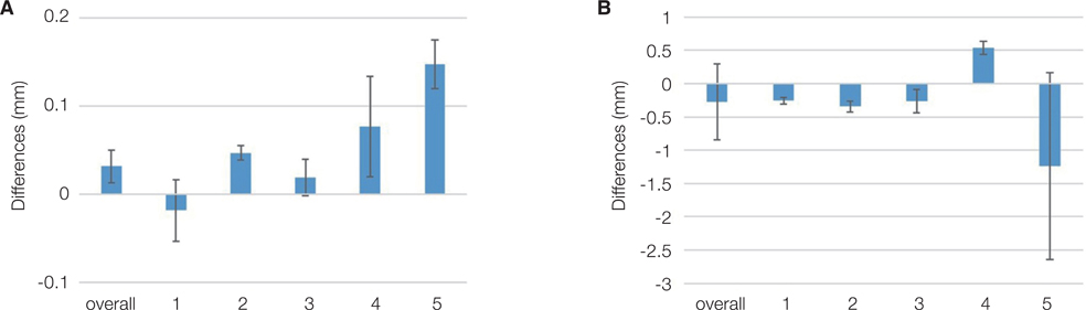
Fig. 4 Results from the comparison between the CI and SI methods; (A) Maxilla: 1-Medial palatine raphe, 2-Hard palate, 3-Residual ridge, 4-Buccal vestibule, 5-Soft palate; (B) Mandible: 1-Residual ridge, 2-Buccal shelf, 3-Retromolar pad, 4-Buccal vestibule, 5-Labial vestibule. None of the values showed statistically significant differences.
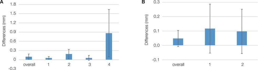
Fig. 5 Results from the comparison between the CI and DI methods; (A) Maxilla: 1-Medial palatine raphe, 2-Hard palate, 3-Residual ridge, 4-Soft palate; (B) Mandible: 1-Residual ridge, 2-Buccal shelf. None of the values showed statistically significant differences.
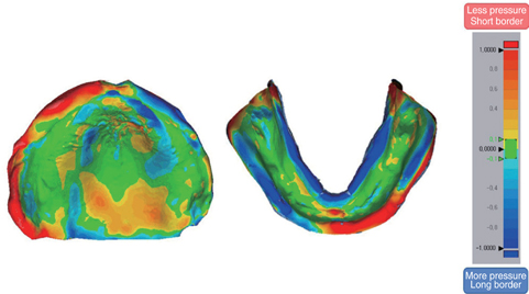
Fig. 6 Color deviation map of superimposition of the areas recorded by the CI and SI methods; ‘Red’ or ‘Yellow’ color means less depressed CI impression technique, and ‘Blue’ color means more depressed CI impression technique. Notice the border of the maxilla (especially left), soft palate, and lingual border of the mandible.
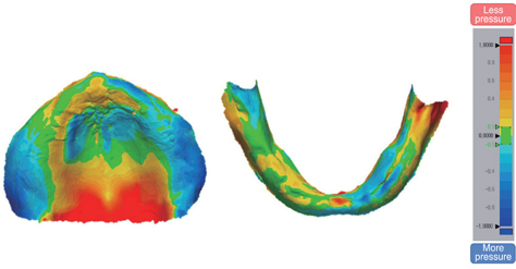
Fig. 7 Color deviation map of superimposition of the areas recorded by the CI and DI methods; ‘Red’ or ‘Yellow’ color means less depressed CI impression technique, and ‘Blue’ color means more depressed CI impression technique. There was a large difference only on the soft palate in the maxilla. That is, the CI method pressed the soft palate less than the DI method.
Reference
-
1. Beuer F, Schweiger J, Edelhoff D. Digital dentistry: an overview of recent developments for CAD/CAM generated restorations. Br Dent J. 2008; 204:505–511.
Article2. Zandinejad A, Lin WS, Atarodi M, Abdel-Azim T, Metz MJ, Morton D. Digital workflow for virtually designing and milling ceramic lithium disilicate veneers: a clinical report. Oper Dent. 2015; 40:241–246.
Article3. Wee AG, Lindsey DT, Kuo S, Johnston WM. Color accuracy of commercial digital cameras for use in dentistry. Dent Mater. 2006; 22:553–559.
Article4. Brennan J. An introduction to digital radiography in dentistry. J Orthod. 2002; 29:66–69.
Article5. Koch GK, Gallucci GO, Lee SJ. Accuracy in the digital workflow: From data acquisition to the digitally milled cast. J Prosthet Dent. 2016; 115:749–754.
Article6. Neumeier TT, Neumeier H. Digital immediate dentures treatment: A clinical report of two patients. J Prosthet Dent. 2016; 116:314–319.
Article7. Alhassani AA, AlGhamdi AS. Inferior alveolar nerve injury in implant dentistry: diagnosis, causes, prevention, and management. J Oral Implantol. 2010; 36:401–407.
Article8. Wenzel A. A review of dentists’ use of digital radiography and caries diagnosis with digital systems. Dentomaxillofac Radiol. 2006; 35:307–314.
Article9. Brawek PK, Wolfart S, Endres L, Kirsten A, Reich S. The clinical accuracy of single crowns exclusively fabricated by digital workflow-the comparison of two systems. Clin Oral Investig. 2013; 17:2119–2125.
Article10. Payer M, Arnetzl V, Kirmeier R, Koller M, Arnetzl G, Jakse N. Immediate provisional restoration of single-piece zirconia implants: a prospective case series - results after 24 months of clinical function. Clin Oral Implants Res. 2013; 24:569–575.11. Joo HS, Park SW, Yun KD, Lim HP. Complete-mouth rehabilitation using a 3D printing technique and the CAD/CAM double scanning method: A clinical report. J Prosthet Dent. 2016; 116:3–7.
Article12. Ohkubo C, Shimpo H, Tokue A, Park EJ, Kim TH. Complete denture fabrication using piezography and CAD-CAM: A clinical report. J Prosthet Dent. 2018; 119:334–338.
Article13. Wimmer T, Gallus K, Eichberger M, Stawarczyk B. Complete denture fabrication supported by CAD/CAM. J Prosthet Dent. 2016; 115:541–546.
Article14. McLaughlin JB, Ramos V Jr. Complete denture fabrication with CAD/CAM record bases. J Prosthet Dent. 2015; 114:493–497.
Article15. AlHelal A, AlRumaih HS, Kattadiyil MT, Baba NZ, Goodacre CJ. Comparison of retention between maxillary milled and conventional denture bases: A clinical study. J Prosthet Dent. 2017; 117:233–238.
Article16. Felton DA, Cooper LF, Scurria MS. Predictable impression procedures for complete dentures. Dent Clin North Am. 1996; 40:39–51.17. Collett HA. Final impressions for complete dentures. J Prosthet Dent. 1970; 23:250–264.
Article18. Goodacre BJ, Goodacre CJ, Baba NZ, Kattadiyil MT. Comparison of denture tooth movement between CADCAM and conventional fabrication techniques. J Prosthet Dent. 2018; 119:108–115.
Article19. Schweiger J, Güth JF, Edelhoff D, Stumbaum J. Virtual evaluation for CAD-CAM-fabricated complete dentures. J Prosthet Dent. 2017; 117:28–33.
Article20. Jacob RF. The traditional therapeutic paradigm: complete denture therapy. J Prosthet Dent. 1998; 79:6–13.
Article21. Ceruti P, Mobilio N, Bellia E, Borracchini A, Catapano S, Gassino G. Simplified edentulous treatment: A multicenter randomized controlled trial to evaluate the timing and clinical outcomes of the technique. J Prosthet Dent. 2017; 118:462–467.
Article22. Kattadiyil MT, AlHelal A, Goodacre BJ. Clinical complications and quality assessments with computer-engineered complete dentures: A systematic review. J Prosthet Dent. 2017; 117:721–728.
Article23. Fang JH, An X, Jeong SM, Choi BH. Development of complete dentures based on digital intraoral impressions-Case report. J Prosthodont Res. 2018; 62:116–120.
Article24. Alsharbaty MHM, Alikhasi M, Zarrati S, Shamshiri AR. A clinical comparative study of 3-dimensional accuracy between digital and conventional implant impression techniques. J Prosthodont. 2019; 28:e902–e908.
Article25. Gan N, Xiong Y, Jiao T. Accuracy of intraoral digital impressions for whole upper jaws, including full dentitions and palatal soft tissues. PLoS One. 2016; 11:e0158800.
Article26. Sharma S, Agarwal S, Sharma D, Kumar S, Glodha N. Impression; Digital vs. conventional: A review. Ann Dent Spec. 2014; 2:9–10.27. Birnbaum NS, Aaronson HB.. virtual becomes reality. Compend Contin Educ Dent. 2008; 29:494496498–505.28. Heo YR, Kim HJ, Son MK, Chung CH. Contour of lingual surface in lower complete denture formed by polished surface impression. J Adv Prosthodont. 2016; 8:472–478.
Article29. Park C, Yang HS, Lim HP, Yun KD, Oh GJ, Park SW. A new fast and simple border molding process for complete dentures using a compound stick gun. Int J Prosthodont. 2016; 29:559–560.
Article30. Dawson PE. Evaluation, diagnosis and treatment of occlusal problems. 3rd ed. St. Louis: Mosby;1989. p. 41–47.31. Collett HA. Complete denture impressions. J Prosthet Dent. 1965; 15:603–614.
Article32. Matsuda T, Goto T, Kurahashi K, Kashiwabara T, Watanabe M, Tomotake Y, Nagao K, Ichikawa T. Digital assessment of preliminary impression accuracy for edentulous jaws: Comparisons of 3-dimensional surfaces between study and working casts. J Prosthodont Res. 2016; 60:206–212.
Article33. Yoon HI, Hwang HJ, Ohkubo C, Han JS, Park EJ. Evaluation of the trueness and tissue surface adaptation of CAD-CAM mandibular denture bases manufactured using digital light processing. J Prosthet Dent. 2018; 120:919–926.
Article34. Pinelli LA, Fais LM, Ricci WA, Reis JM. In vitro comparisons of casting retention on implant abutments among commercially available and experimental castor oil-containing dental luting agents. J Prosthet Dent. 2013; 109:319–324.
Article35. Koller MM, Merlini L, Spandre G, Palla S. A comparative study of two methods for the orientation of the occlusal plane and the determination of the vertical dimension of occlusion in edentulous patients. J Oral Rehabil. 1992; 19:413–425.
Article36. Kawai Y, Murakami H, Shariati B, Klemetti E, Blomfield JV, Billette L, Lund JP, Feine JS. Do traditional techniques produce better conventional complete dentures than simplified techniques? J Dent. 2005; 33:659–668.
Article37. Chaware SH, Fernandes F. Tissue stress evaluation at border seal area using patient-manipulated custom tray-modified closed-mouth functional technique for flat mandibular ridges. J Int Oral Health. 2018; 10:77–82.
Article38. Azzam MK, Yurkstas AA, Kronman J. The sublingual crescent extension and its relation to the stability and retention of mandibular complete dentures. J Prosthet Dent. 1992; 67:205–210.
Article39. Kim JE, Amelya A, Shin Y, Shim JS. Accuracy of intraoral digital impressions using an artificial landmark. J Prosthet Dent. 2017; 117:755–761.40. Logozzo S, Zanetti EM, Franceschini G, Kilpela A, Makynen A. Recent advances in dental optics - Part I: 3D intraoral scanners for restorative dentistry. Opt Lasers Eng. 2014; 54:203–221.
Article41. Patzelt SB, Bishti S, Stampf S, Att W. Accuracy of computeraided design/computer-aided manufacturing-generated dental casts based on intraoral scanner data. J Am Dent Assoc. 2014; 145:1133–1140.
Article42. Flügge TV, Schlager S, Nelson K, Nahles S, Metzger MC. Precision of intraoral digital dental impressions with iTero and extraoral digitization with the iTero and a model scanner. Am J Orthod Dentofacial Orthop. 2013; 144:471–478.
Article43. Ender A, Mehl A. Accuracy of complete-arch dental impressions: a new method of measuring trueness and precision. J Prosthet Dent. 2013; 109:121–128.
Article44. Patzelt SB, Emmanouilidi A, Stampf S, Strub JR, Att W. Accuracy of full-arch scans using intraoral scanners. Clin Oral Investig. 2014; 18:1687–1694.
Article
- Full Text Links
-
- Actions
-
Cited
- CITED
-
- Close
- Share
-
- Similar articles
-
- Three-dimensional analysis of the normal dentition and edentulous maxilla of Koreans
- Comparison of denture supporting area and retention between different mandibular denture impression techniques in a fully edentulous patient with severe alveolar bone resorption: a case report
- Comparison of intraoral scanning and conventional impression techniques using 3-dimensional superimposition
- Fabrication of complete denture using Centric tray and closed mouth technique for edentulous patient
- Complete denture made with closed-mouth impression technique on severely atrophied edentulous jaw
