J Dent Rehabil Appl Sci.
2024 Nov;40(4):268-278. 10.14368/jdras.2024.40.4.268.
Comparison of denture supporting area and retention between different mandibular denture impression techniques in a fully edentulous patient with severe alveolar bone resorption: a case report
- Affiliations
-
- 1Division of Prosthodontics, Department of Dentistry, Asan Medical Center, Seoul, Republic of Korea
- 2Division of Prosthodontics, Department of Dentistry, Asan Medical Center, College of Medicine, University of Ulsan, Seoul, Republic of Korea
- KMID: 2562771
- DOI: http://doi.org/10.14368/jdras.2024.40.4.268
Abstract
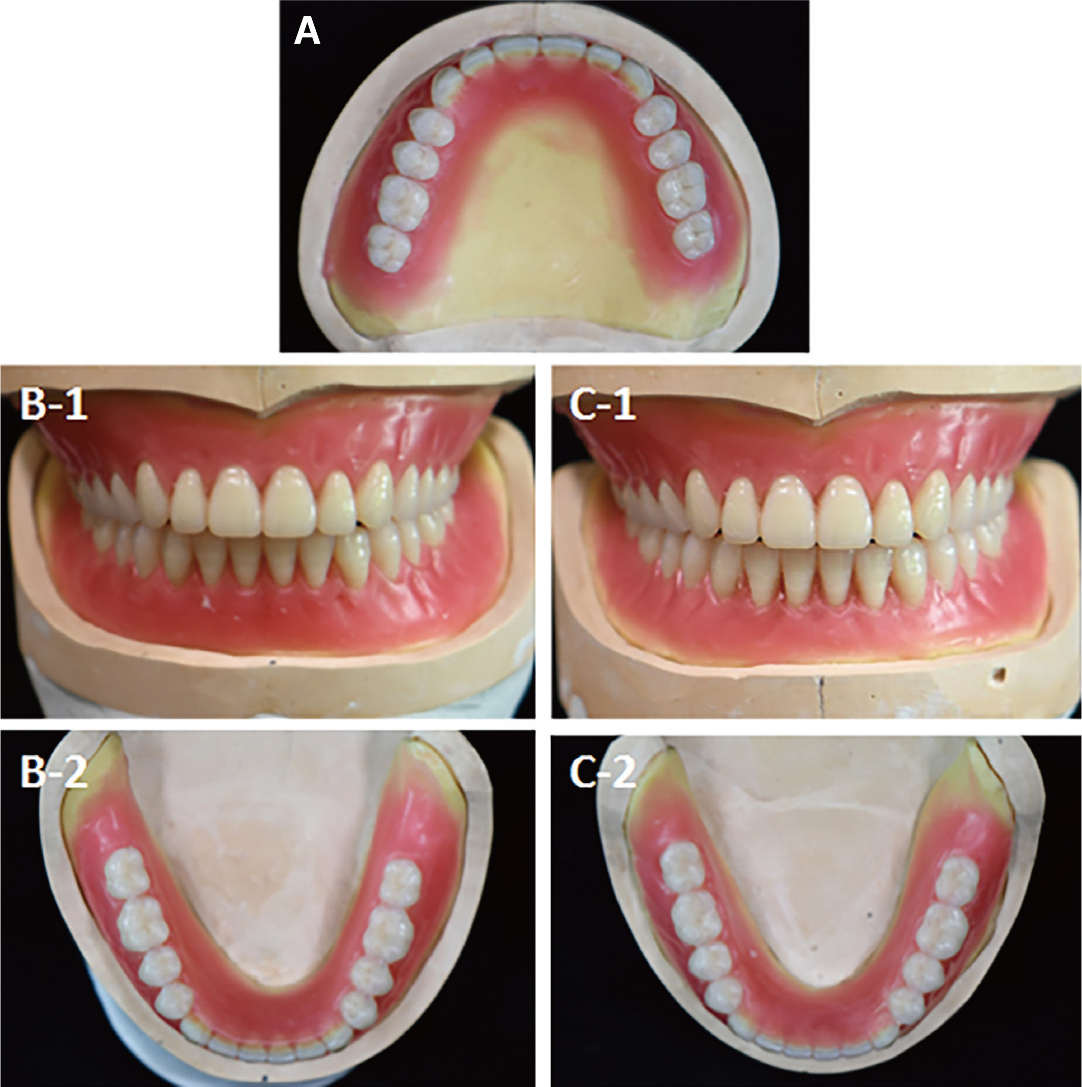
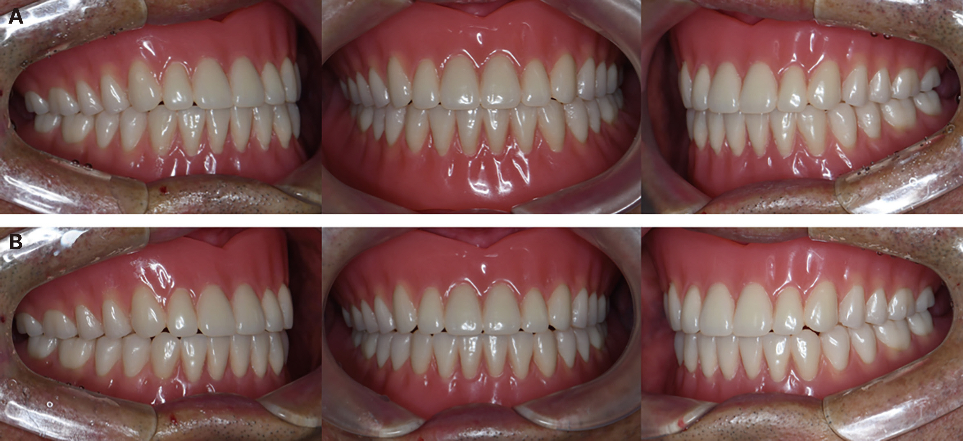
- In fully edentulous patients with severe mandibular alveolar bone resorption, the retention and stability of a mandibular complete denture may be limited. There are two primary impression techniques for complete denture fabrication: the open-mouth impres-sion technique and the closed-mouth impression technique. These two methods differ in how they achieve retention and stability, and each has its own advantages and disadvantages. The study aims to compare the difference in supporting area, retention, and patient satisfaction between mandibular complete dentures fabricated using two impression techniques: the open-mouth impres-sion technique and the closed-mouth impression technique.
Keyword
Figure
Reference
-
References
1. Zarb GA, Bolender CL, Eckert SE, Fenton AH, Jacob RF, Mericske-Stern R. Prosthodontic treatment for edentulous patients: Complete dentures and implant-supported prestheses. 12th ed. St. Louis: Mosby;2004. p. 24–33. p. 73–99.2. Collett HA. 1970; Final impressions for complete dentures. J Prosthet Dent. 23:250–64. DOI: 10.1016/0022-3913(70)90180-0. PMID: 4905179.3. Abe J, Kokubo K, Sato K. Mandibular Suction-effective denture and BPS: A complete guide. Tokyo: Quintessence Publishing Co. Ltd.;2012. p. 46–78.4. Cho IH, Kwon KR, Kwon HB, Kim MH, Lee CH, Jeong CM, Park CJ, Park SW, Lee JH, Cho HW, Kim HJ, Moon HS, Chung MK, Chung CH, Lim YJ, Lee JS, Song KW, Han CH. Prosthodontic treatment for edentulous patients. 2nd ed. Seoul: Dental Wisdom;2014. p. 98–145.5. Sato K. What is Suction Denture? Tokyo: Dental Diamond Co.;2014. p. 32–57.6. Jassim TK, Kareem AE, Alloaibi MA. 2020; In vivo evaluation of the impact of various border molding materials and techniques on the retention of complete maxillary denture. Dent Med Probl. 57:191–6. DOI: 10.17219/dmp/115104. PMID: 32649808.7. Epifania E, Sanzullo R, Sorrentino R, Ausiello P. 2018; Evaluation of Satisfaction Perceived by Prosthetic Patients Compared to Clinical and Technical Variables. J Int Soc Prev Community Dent. 8:252–8. DOI: 10.4103/jispcd.JISPCD_27_18. PMID: 29911064. PMCID: PMC5985683.8. Abe J. 2010; Difference of preliminary impression takings between conventional mandibular complete denture and the mandibular complete denture intended with effective suction. Pract Prosthodont. 43:510–24.9. Tallgren A. 1972; The continuing reduction of the residual alveolar ridges in complete denture wearers: a mixed-longitudinal study covering 25 years. J Prosthet Dent. 27:120–32. DOI: 10.1016/0022-3913(72)90188-6. PMID: 4500507.
- Full Text Links
- Actions
-
Cited
- CITED
-
- Close
- Share
- Similar articles
-
- Complete denture fabricated by Jiro Abe's method for edentulous patient with severe alveolar ridge resorption: a case report
- Fabrication of complete denture using Centric tray and closed mouth technique for edentulous patient
- Complete denture made with closed-mouth impression technique on severely atrophied edentulous jaw
- Complete denture fabrication of edentulous patient withsevere alveolar bone resorption using suction mechanism: A case report
- Complete denture rehabilitation of edentulous patient with severe alveolar bone resorption andcondyle fracture using gothic arch tracing and closed mouth impression technique: A case report


