Flow Cytometric Analysis of T Cells in Hemophagocytic Lymphohistiocytosis
- Affiliations
-
- 1Department of Laboratory Medicine and Genetics, Samsung Medical Center, Sungkyunkwan University School of Medicine, Seoul, Korea. duck.cho@samsung.com
- 2Division of Hematology and Oncology, Department of Medicine, Samsung Medical Center, Sungkyunkwan University School of Medicine, Seoul, Korea. kstwoh@skku.edu
- KMID: 2450965
- DOI: http://doi.org/10.3343/alm.2019.39.5.430
Abstract
- BACKGROUND
T cell immunophenotypes in patients with hemophagocytic lymphohistiocytosis (HLH) have been described. Downregulation of CD5 or CD7 on T cells has been reported in patients with Epstein-Barr virus (EBV)-positive HLH. As the utility of T cell immunophenotypes as an adjunctive diagnostic or a prognostic marker for HLH has not been evaluated, we analyzed T cell immunophenotypes in HLH patients for this purpose.
METHODS
We classified 45 HLH patients into three subgroups: EBV-positive HLH (N=27), EBV-negative secondary HLH (N=15), and familial HLH (N=3). We retrospectively characterized downregulation patterns of CD5 or CD7 on activated T cells, using flow cytometry. Overall survival was estimated using Kaplan-Meier curves and compared using the log-rank test.
RESULTS
An aberrant immunophenotype, including CD5 and/or CD7 downregulation on T cells, was observed in 55.6% (15/27) of the EBV-positive HLH patients and 100% of the familial HLH (3/3). Only one (1/15, 6.7%) patient with EBV-negative secondary HLH showed an aberrant loss of CD7 antigen on CD8+ T cells. The presence of an aberrant T cell immunophenotype was not related to overall survival in EBV-positive HLH and EBV-negative secondary HLH patients.
CONCLUSIONS
An aberrant T cell immunophenotype may assist in discriminating EBV-negative secondary HLH and EBV-positive HLH. However, it may not be useful as a prognostic marker.
Keyword
MeSH Terms
Figure
-
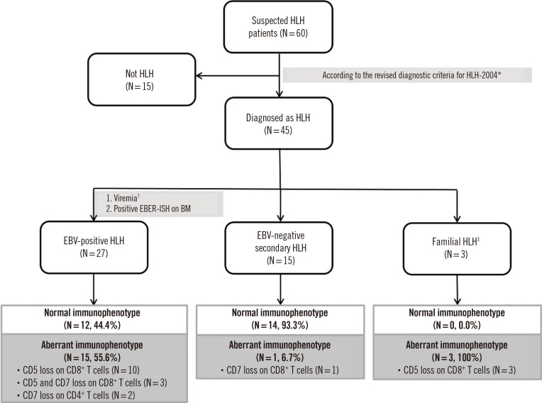
Fig. 1 Study design flowchart for classification of HLH patients.*HLH patients were identified according to the 2004 criteria [13]; †>2,000 genome copies/mL of serum; ‡Three familial HLH cases were diagnosed as type 3 familial HLH based on detection of pathogenic variants of the UNC13D gene.Abbreviations: HLH, hemophagocytic lymphohistiocytosis; EBV, Epstein-Barr virus; EBER-ISH, Epstein-Barr encoding region in situ hybridization; BM, bone marrow; MDS, myelodysplastic syndromes; LPD, lymphoproliferative disease.
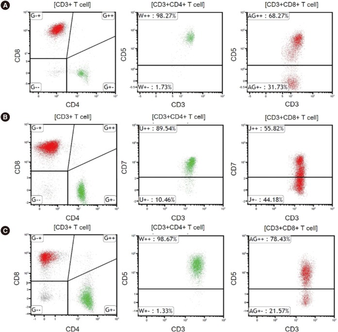
Fig. 2 Representative flow cytometric findings in HLH patients. Red and green colors indicate CD8+ T cells and CD4+ T cells, respectively.(A) CD5 downregulation on CD8+ T cells in EBV-positive HLH. (B) CD7 downregulation on CD8+ T cells in EBV-negative secondary HLH. (C) CD5 downregulation on CD8+ T cells in familial HLH.Abbreviations: HLH, hemophagocytic lymphohistiocytosis; EBV, Epstein-Barr virus.
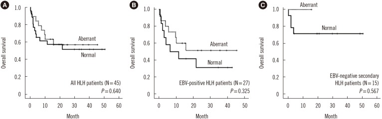
Fig. 3 Kaplan-Meier survival analysis of HLH patients based on the presence of an aberrant T cell immunophenotype. (A) Overall survival of all HLH patients. (B) Overall survival of EBV-positive HLH patients. (C) Overall survival of EBV-negative secondary HLH patients.Abbreviations: HLH, hemophagocytic lymphohistiocytosis; EBV, Epstein-Barr virus.
Reference
-
1. Otrock ZK, Eby CS. Clinical characteristics, prognostic factors, and outcomes of adult patients with hemophagocytic lymphohistiocytosis. Am J Hematol. 2015; 90:220–224. PMID: 25469675.2. McCall CM, Mudali S, Arceci RJ, Small D, Fuller S, Gocke CD, et al. Flow cytometric findings in hemophagocytic lymphohistiocytosis. Am J Clin Pathol. 2012; 137:786–794. PMID: 22523218.3. Shin SY, Lee K, Jang MA, Lee ST, Yoo KH, Koo HH, et al. First report on familial hemophagocytic lymphohistiocytosis with an abnormal immunophenotype and T cell monoclonality in Korea. Ann Lab Med. 2015; 35:155–158. PMID: 25553300.4. Wada T, Sakakibara Y, Nishimura R, Toma T, Ueno Y, Horita S, et al. Down-regulation of CD5 expression on activated CD8+ T cells in familial hemophagocytic lymphohistiocytosis with perforin gene mutations. Hum Immunol. 2013; 74:1579–1585. PMID: 24051121.5. Henter JI, Horne A, Arico M, Egeler RM, Filipovich AH, Imashuku S, et al. HLH-2004: Diagnostic and therapeutic guidelines for hemophagocytic lymphohistiocytosis. Pediatr Blood Cancer. 2007; 48:124–131. PMID: 16937360.6. Stevens SJ, Pronk I, Middeldorp JM. Toward standardization of Epstein-Barr virus DNA load monitoring: unfractionated whole blood as preferred clinical specimen. J Clin Microbiol. 2001; 39:1211–1216. PMID: 11283029.7. Jamal S, Picker LJ, Aquino DB, McKenna RW, Dawson DB, Kroft SH. Immunophenotypic analysis of peripheral T-cell neoplasms. A multiparameter flow cytometric approach. Am J Clin Pathol. 2001; 116:512–526. PMID: 11601136.8. Brisse E, Wouters CH, Matthys P. Advances in the pathogenesis of primary and secondary haemophagocytic lymphohistiocytosis: differences and similarities. Br J Haematol. 2016; 174:203–217. PMID: 27264204.9. Ishii E. Hemophagocytic lymphohistiocytosis in children: pathogenesis and treatment. Front Pediatr. 2016; 4:47. PMID: 27242976.10. Sieni E, Cetica V, Mastrodicasa E, Pende D, Moretta L, Griffiths G, et al. Familial hemophagocytic lymphohistiocytosis: a model for understanding the human machinery of cellular cytotoxicity. Cell Mol Life Sci. 2012; 69:29–40. PMID: 21990010.11. Sieni E, Cetica V, Hackmann Y, Coniglio ML, Da Ros M, Ciambotti B, et al. Familial hemophagocytic lymphohistiocytosis: when rare diseases shed light on immune system functioning. Front Immunol. 2014; 5:167. PMID: 24795715.12. Yoon HS, Kim HJ, Yoo KH, Sung KW, Koo HH, Kang HJ, et al. UNC13D is the predominant causative gene with recurrent splicing mutations in Korean patients with familial hemophagocytic lymphohistiocytosis. Haematologica. 2010; 95:622–626. PMID: 20015888.13. Koh KN, Im HJ, Chung NG, Cho B, Kang HJ, Shin HY, et al. Clinical features, genetics, and outcome of pediatric patients with hemophagocytic lymphohistiocytosis in Korea: report of a nationwide survey from Korea Histiocytosis Working Party. Eur J Haematol. 2015; 94:51–59. PMID: 24935083.14. Manji F, Wilson E, Mahe E, Gill J, Conly J. Acute HIV infection presenting as hemophagocytic lymphohistiocytosis: case report and review of the literature. BMC Infect Dis. 2017; 17:633. PMID: 28931369.15. Brisse E, Wouters CH, Andrei G, Matthys P. How viruses contribute to the pathogenesis of hemophagocytic lymphohistiocytosis. Front Immunol. 2017; 8:1102. PMID: 28936212.16. Marsh RA. Epstein-Barr virus and hemophagocytic lymphohistiocytosis. Front Immunol. 2017; 8:1902. PMID: 29358936.17. Ishii E, Ohga S, Imashuku S, Yasukawa M, Tsuda H, Miura I, et al. Nationwide survey of hemophagocytic lymphohistiocytosis in Japan. Int J Hematol. 2007; 86:58–65. PMID: 17675268.18. Imashuku S. Clinical features and treatment strategies of Epstein-Barr virus-associated hemophagocytic lymphohistiocytosis. Crit Rev Oncol Hematol. 2002; 44:259–272. PMID: 12467966.19. Kawa K. Epstein-Barr virus--associated diseases in humans. Int J Hematol. 2000; 71:108–117. PMID: 10745621.20. Dojcinov SD, Fend F, Quintanilla-Martinez L. EBV-positive lymphoproliferations of B- T- and NK-cell derivation in non-immunocompromised hosts. Pathogens. 2018; 7.21. Lin MT, Chang HM, Huang CJ, Chen WL, Lin CY, Lin CY, et al. Massive expansion of EBV+ monoclonal T cells with CD5 down regulation in EBV-associated haemophagocytic lymphohistiocytosis. J Clin Pathol. 2007; 60:101–103. PMID: 17213357.22. Weisberger J, Cornfield D, Gorczyca W, Liu Z. Down-regulation of pan-T-cell antigens, particularly CD7, in acute infectious mononucleosis. Am J Clin Pathol. 2003; 120:49–55. PMID: 12866372.23. Wada T, Kurokawa T, Toma T, Shibata F, Tone Y, Hashida Y, et al. Immunophenotypic analysis of Epstein-Barr virus (EBV)-infected CD8(+) T cells in a patient with EBV-associated hemophagocytic lymphohistiocytosis. Eur J Haematol. 2007; 79:72–75. PMID: 17532761.24. Karandikar NJ, Kroft SH, Yegappan S, Rogers BB, Aquino VM, Lee KM, et al. Unusual immunophenotype of CD8+ T cells in familial hemophagocytic lymphohistiocytosis. Blood. 2004; 104:2007–2009. PMID: 15205266.25. Toga A, Wada T, Sakakibara Y, Mase S, Araki R, Tone Y, et al. Clinical significance of cloned expansion and CD5 down-regulation in Epstein-Barr Virus (EBV)-infected CD8+ T lymphocytes in EBV-associated hemophagocytic lymphohistiocytosis. J Infect Dis. 2010; 201:1923–1932. PMID: 20443735.26. Manno EC, Salfa I, Palma P, Bertaina A, Lombardi A, Moretta F, et al. Familial hemophagocytic lymphohistiocytosis type 3 diagnosed at school age: a case report. J Pediatr Hematol Oncol. 2014; 36:e128–e130. PMID: 23669735.27. Tembhare P, Yuan CM, Xi L, Morris JC, Liewehr D, Venzon D, et al. Flow cytometric immunophenotypic assessment of T-cell clonality by Vbeta repertoire analysis: detection of T-cell clonality at diagnosis and monitoring of minimal residual disease following therapy. Am J Clin Pathol. 2011; 135:890–900. PMID: 21571962.28. Mehta RS. Hemophagocytic lymphohistiocytosis (HLH): a review of literature. Medical Oncology. 2013; 30:740. PMID: 24105023.29. Imashuku S, Teramura T, Tauchi H, Ishida Y, Otoh Y, Sawada M, et al. Longitudinal follow-up of patients with Epstein-Barr virus-associated hemophagocytic lymphohistiocytosis. Haematologica. 2004; 89:183–188. PMID: 15003893.30. Ohga S, Kudo K, Ishii E, Honjo S, Morimoto A, Osugi Y, et al. Hematopoietic stem cell transplantation for familial hemophagocytic lymphohistiocytosis and Epstein-Barr virus-associated hemophagocytic lymphohistiocytosis in Japan. Pediatr Blood Cancer. 2010; 54:299–306. PMID: 19827139.
- Full Text Links
-
- Actions
-
Cited
- CITED
-
- Close
- Share
-
- Similar articles
-
- Two Cases of Hemophagocytic Lymphohistiocytosis Following Kikuchi's Disease
- Multiple Ecthyma Gangrenosum in a Hemophagocytic Lymphohistiocytosis Patient
- A Case of Secondary Precursor B-cell Acute Lymphoblastic Leukemia Occurring after Treatment of Hemophagocytic Lymphohistiocytosis
- A Case of Hemophagocytic Lymphohistiocytosis Presenting with Neck Mass in a Child
- Hemophagocytic lymphohistiocytosis secondary to histoplasmosis
