Common conditions associated with mandibular canal widening: A literature review
- Affiliations
-
- 1Department of Oral Medicine, School of Dentistry, Shahid Beheshti University of Medical Sciences, Tehran, Iran.
- 2Department of Oral and Maxillofacial Radiology, School of Dentistry, Shahid Beheshti University of Medical Sciences, Tehran, Iran.
- 3Department of Orthodontic, School of Dentistry, Shahid Beheshti University of Medical Sciences, Tehran, Iran. behnaz1357@yahoo.com
- 4School of Dentistry, Shahid Beheshti University of Medical Sciences, Tehran, Iran.
- KMID: 2450174
- DOI: http://doi.org/10.5624/isd.2019.49.2.87
Abstract
- PURPOSE
The aim of this study was to review the common conditions associated with mandibular canal widening.
MATERIALS AND METHODS
General search engines and specialized databases including Google Scholar, PubMed, PubMed Central, Science Direct, and Scopus were used to find relevant studies by using the following keywords: "mandibular canal,""alveolar canal,""inferior alveolar nerve canal,""inferior dental canal,""inferior mandibular canal,""widening,""enlargement,""distension,""expansion," and "dilation."
RESULTS
In total, 130 articles were found, of which 80 were broadly relevant to the topic. We ultimately included 38 articles that were closely related to the topic of interest. When the data were compiled, the following 7 lesions were found to have a relationship with mandibular canal widening: non-Hodgkin lymphoma, osteosarcoma, schwannoma, neurofibroma, vascular malformation/hemangioma, multiple endocrine neoplasia syndromes, and perineural spreading or invasion.
CONCLUSION
When clinicians encounter a lesion associated with mandibular canal widening, they should immediately consider these entities in the differential diagnosis. Doing so will help dentists make more accurate diagnoses and develop better treatment plans based on patients' radiographs.
MeSH Terms
Figure
-

Fig. 1 Panoramic reconstructed and 3-dimensional cone-beam computed tomographic images show mandibular canal and mental foramen widening in a patient with non-Hodgkin lymphoma, respectively.
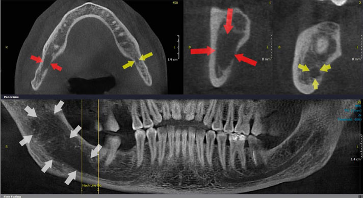
Fig. 2 Cone-beam computed tomographic images show mandibular canal widening in a patient with osteosarcoma (arrows show the normal canal and mandibular canal widening).
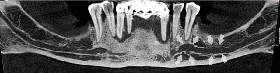
Fig. 3 A panoramic reconstructed cone-beam computed tomographic image shows mandibular canal widening in a patient with schwannoma.
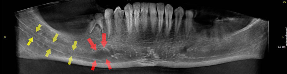
Fig. 4 A panoramic reconstructed cone-beam computed tomographic image shows mandibular canal widening in a patient with neurofibroma (arrows show the normal canal and mandibular canal widening).

Fig. 5 A panoramic image shows mandibular canal widening in a patient with central hemangioma.
Reference
-
1. Ai CJ, Jabar NA, Lan TH, Ramli R. Mandibular canal enlargement: clinical and radiological characteristics. J Clin Imaging Sci. 2017; 7:28.2. Abdi I, Taheri Talesh K, Yazdani J, Keshavarz Meshkin Fam S, Ghavimi MA, Arta SA. The effect of ameloblastoma and keratocystic odontogenic tumor on the displacement pattern of inferior alveolar canal in CBCT examinations. J Dent Res Dent Clin Dent Prospects. 2016; 10:155–161.
Article3. Terzic A, Becker M, Imholz B, Scollozi P. Unilateral widening of the inferior alveolar nerve canal: a rare anatomic variant mimicking disease. Oral Radiol. 2013; 29:160–165.
Article4. Guimarães DM, Pontes FS, Da Mata Rezende Ddos S, Pontes HA. Anatomical variation of mandibular canal simulating a recurrence of odontogenic tumor. Ann Maxillofac Surg. 2014; 4:107–109.
Article5. Jung YH, Cho BH. Radiographic evaluation of the course and visibility of the mandibular canal. Imaging Sci Dent. 2014; 44:273–278.
Article6. Munhoz L, Marsan FP, Arita ES. Radiographic enlargement of mandibular canal as an extranodal primary non-Hodgkin's lymphoma early sign in an asymptomatic patient. Case Rep Dent. 2017; 2017:9193165.
Article7. Mortazavi H, Baharvand M. Review of common conditions associated with periodontal ligament widening. Imaging Sci Dent. 2016; 46:229–237.
Article8. Mojaver YN, Sahebjamie M, Tirgary F, Eslami M, Rezvani G. Enlargement of mandibular canal with tongue paresthesia caused by extranodal B-cell lymphoma: a case report. Oral Oncol Extra. 2005; 41:97–99.
Article9. Buric N, Jovanovic G, Radovanovic Z, Buric M, Tijanic M. Radiographic enlargement of mandibular canal as first feature of non-Hodgkin's lymphoma. Dentomaxillofac Radiol. 2010; 39:383–388.10. Bertolotto M, Cecchini G, Martinoli C, Perrone R, Garlaschi G. Primary lymphoma of the mandible with diffuse widening of the mandibular canal: report of a case. Eur Radiol. 1996; 6:637–639.
Article11. Barber HD, Stewart JC, Baxter WD. Non-Hodgkin's lymphoma involving the inferior alveolar canal and mental foramen: report of a case. J Oral Maxillofac Surg. 1992; 50:1334–1336.12. Chittaranjan B, Tejasvi MA, Babu BB, Geetha P. Intramedullary osteosarcoma of the mandible: a clinicoradiologic perspective. J Clin Imaging Sci. 2014; 4:Suppl 2. 6.
Article13. Yagan R, Radivoyevitch M, Bellon EM. Involvement of the mandibular canal: early sign of osteogenic sarcoma of the mandible. Oral Surg Oral Med Oral Pathol. 1985; 60:56–60.
Article14. Givol N, Buchner A, Taicher S, Kaffe I. Radiological features of osteogenic sarcoma of the jaws. A comparative study of different radiographic modalities. Dentomaxillofac Radiol. 1998; 27:313–320.
Article15. Garrington GE, Scofield HH, Cornyn J, Hooker SP. Osteosarcoma of the jaws. Analysis of 56 cases. Cancer. 1967; 20:377–391.16. Abouchadi A, Guerrouani A, Ribag Y, El Khatib K, Nassih M. Intrabony schwannoma of the mandible: case report and review of literature. Open J Stomatol. 2014; 4:233–237.
Article17. Minić AJ. Central schwannoma of the maxilla. Int J Oral Maxillofac Surg. 1992; 21:297–298.
Article18. Vartiainen VM, Siponen M, Salo T, Rosberg J, Apaja-Sarkkinen M. Widening of the inferior alveolar canal: a case report with atypical lymphocytic infiltration of the nerve. Oral Surg Oral Med Oral Pathol Oral Radiol Endod. 2008; 106:e35–e39.
Article19. Redman RS, Guccion JG, Spector CJ, Keegan BP. Cellular schwannoma of the mandible: a case report with ultrastructural and immunohistochemical observations. J Oral Maxillofac Surg. 1996; 54:339–344.
Article20. Chi AC, Carey J, Muller S. Intraosseous schwannoma of the mandible: a case report and review of the literature. Oral Surg Oral Med Oral Pathol Oral Radiol Endod. 2003; 96:54–65.
Article21. Khan M, Ohri N. Oral manifestations of Type I neurofibromatosis in a family. J Clin Exp Dent. 2011; 3:e483–e486.
Article22. Mortazavi H, Safi Y, Baharvand M, Rahmani S, Jafari S. Peripheral exophytic oral lesions: a clinical decision tree. Int J Dent. 2017; 2017:9193831.
Article23. Dalili Z, Adham G. Intraosseous neurofibroma and concurrent involvement of the mandible, maxilla and orbit: report of a case. Iran J Radiol. 2012; 9:45–49.
Article24. Visnapuu V, Peltonen S, Tammisalo T, Peltonen J, Happonen RP. Radiographic findings in the jaws of patients with neurofibromatosis 1. J Oral Maxillofac Surg. 2012; 70:1351–1357.
Article25. González-Arriagada WA, Dias MA, Dias PD, Martínez-Martínez M, Sena-Filho M, de Almeida OP. Oral encapsulated vascular malformation: an undescribed presentation in the mouth. J Clin Exp Dent. 2016; 8:e84–e88.
Article26. Siu WW, Weill A, Gariepy JL, Moret J, Marotta T. Arteriovenous malformation of the mandible: embolization and direct injection therapy. J Vasc Interv Radiol. 2001; 12:1095–1098.
Article27. Sakkas N, Schramm A, Metzger MC, Berlis A, Schmelzeisen R, Otten JE, et al. Arteriovenous malformation of the mandible: a life-threatening situation. Ann Hematol. 2007; 86:409–413.
Article28. Gómez Oliveira G, García-Rozado A, Luaces Rey R. Intraosseous mandibular hemangioma. A case report and review of the literature. Med Oral Patol Oral Cir Bucal. 2008; 13:E496–E498.29. Ozdemir R, Alagoz S, Uysal AC, Unlu RE, Ortak T, Sensoz O. Intraosseous hemangioma of the mandible: a case report and review of the literature. J Craniofac Surg. 2002; 13:38–43.30. Vaezeafshar R, Liu SY, Sidell D. Inferior alveolar nerve hemangioma. Laryngoscope. 2016; 126:2168–2170.
Article31. Accurso B, Mercado A, Allen CM. Multiple endocrine neoplasia-2B presenting with orthodontic relapse. Angle Orthod. 2010; 80:585–590.
Article32. Kahn MA, Cote GJ, Gagel RF. RET protooncogene mutational analysis in multiple endocrine neoplasia syndrome type 2B: case report and review of the literature. Oral Surg Oral Med Oral Pathol Oral Radiol Endod. 1996; 82:288–294.33. Iihara M, Yamashita T, Okamoto T, Kanbe M, Yamazaki K, Egawa S, et al. A nationwide clinical survey of patients with multiple endocrine neoplasia type 2 and familial medullary thyroid carcinoma in Japan. Jpn J Clin Oncol. 1997; 27:128–134.
Article34. Schenberg ME, Zajac JD, Lim-Tio S, Collier NA, Brooks AM, Reade PC. Multiple endocrine neoplasia syndrome - type 2b. Case report and review. Int J Oral Maxillofac Surg. 1992; 21:110–114.35. Sundar GT, Sherigar V, Shetty SS, Satya S, Gohil SM. Mandibular canal widening and bell's palsy: sequelae of perineural invasion in oral cancer. Case Rep Dent. 2016; 2016:3010934.
Article36. Cox CS, Stallworth DG, Ahmed KA, Wadsworth JT, Wenig B, Chung CH, et al. Perineural tumor spread involving the trigeminal and facial nerves: a review of critical imaging findings. Ann Otolaryngol Rhinol. 2017; 4:1177.37. Laske RD, Scholz I, Ikenberg K, Meerwein C, Vital DG, Studer G, et al. Perineural invasion in squamous cell carcinoma of the oral cavity: histology, tumor stage, and outcome. Laryngoscope Investig Otolaryngol. 2016; 1:13–18.
Article38. Dantas AN, Morais EF, Macedo RA, Tinôco JM, Morais Mde L. Clinicopathological characteristics and perineural invasion in adenoid cystic carcinoma: a systematic review. Braz J Otorhinolaryngol. 2015; 81:329–335.
Article39. Shamim T, Varghese VI, Shameena PM, Sudha S. Primary intraosseous adenoid cystic carcinoma of the mandible with lung metastasis: a case report. J Oral Sci. 2008; 50:95–98.
Article40. Koivisto T, Chiona D, Milroy LL, McClanahan SB, Ahmad M, Bowles WR. Mandibular canal location: cone-beam computed tomography examination. J Endod. 2016; 42:1018–1021.
Article41. Matzko J, Becker DG, Phillips CD. Obliteration of fat planes by perineural spread of squamous cell carcinoma along the inferior alveolar nerve. AJNR Am J Neuroradiol. 1994; 15:1843–1845.42. Bagatin M, Orihovac Z, Mohammed AM. Perineural invasion by carcinoma of the lower lip. J Craniomaxillofac Surg. 1995; 23:155–159.
Article43. Campos MS, Fontes A, Marocchio LS, Nunes FD, de Sousa SC. Clinicopathologic and immunohistochemical features of oral neurofibroma. Acta Odontol Scand. 2012; 70:577–582.
Article44. Naikmasur VG, Sattur AP, Burde K, Nandimath K, Thakur AR. Central hemangioma of the mandible: role of imaging in evaluation. Oral Radiol. 2010; 26:46–51.
Article
- Full Text Links
-
- Actions
-
Cited
- CITED
-
- Close
- Share
-
- Similar articles
-
- Biomechanics in various mandibular widening procedures
- Analysis and evaluation of relative positions of mandibular third molar and mandibular canal impacts
- The clinical study of the mandibular canal location in mandibular molar areas using dentascan
- A retrospective study on incidence of C-shaped canals in mandibular second molars
- Root canal treatment of a mandibular second premolar with three separate root canals
