Applications of digital subtraction angiography in the management of penetrating injuries of the maxillofacial region: A case report
- Affiliations
-
- 1Department of Oral and Maxillofacial Surgery, School of Dentistry, Federal University of Minas Gerais, Belo Horizonte, Minas Gerais, Brazil. wagnerhcastro@hotmail.com
- 2Department of Head and Neck Surgery, School of Medicine, Federal University of Minas Gerais, Belo Horizonte, Minas Gerais, Brazil.
- KMID: 2450171
- DOI: http://doi.org/10.5624/isd.2018.48.4.295
Abstract
- This report presents a clinical case of trauma due to assault with a knife, and describes the importance of using the correct imaging modality in cases of facial penetrating trauma involving the superficial and deep anatomical planes. Penetrating wounds in the maxillofacial region are rare and poorly reported, but can result in serious complications that are difficult to resolve and may compromise the patient's quality of life, especially when large blood vessels or other vital structures are involved. Thus, it is essential to determine the extent of the affected blood vessels and the proximity of the retained object to the anatomical structures. In this case, digital subtraction angiography was the imaging modality chosen. The use of appropriate imaging examinations allows a proper map of the surgical field, reducing the chances of vascular damage during the surgical procedure.
MeSH Terms
Figure
-
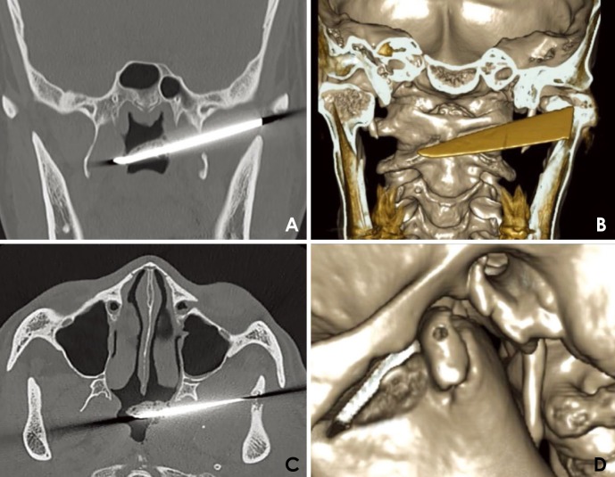
Fig. 1 Preoperative computed tomographic images. A. A coronal image shows the knife fragment in the region of the infratemporal fossa and nasopharynx. B. An axial image shows the knife fragment in the region of the infratemporal fossa and nasopharynx. C. A 3-dimensional reconstructed image reveals the knife path. D. A 3-dimensional reconstruction demonstrates the fracture of the left condylar head.
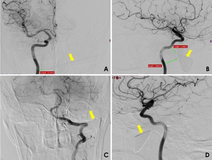
Fig. 2 Selective digital subtraction angiography images of the internal carotid arteries show the penetrating object, indicated with yellow arrows. A. Anteroposterior view of the right internal carotid artery. B. Lateral view of the right internal carotid artery. C. Posteroanterior view of the left internal carotid artery. D. Lateral view of the carotid artery.
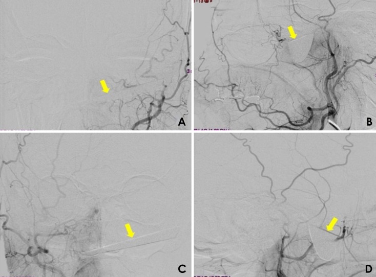
Fig. 3 Selective digital subtraction angiography images of the external carotid arteries show the penetrating object, indicated with yellow arrows. A. Anteroposterior view of the right external carotid artery. B. Lateral view of the right external carotid artery. C. Anteroposterior view of the left external carotid artery. D. Lateral view of the left external carotid artery.
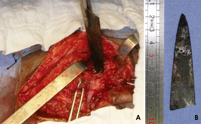
Fig. 4 Surgical procedure. A. Surgical access performed to remove the penetrating object. B. The removed fragment of the knife.
Reference
-
1. Carneiro JT Jr, da Silva Tabosa AK, de Souza FJ Jr, Shinohara EH. Orbitoethmoidal impacted injury by kitchen knife causing abducens nerve palsy. Oral Maxillofac Surg. 2011; 15:107–108. PMID: 20336336.
Article2. Shinohara EH, Heringer L, de Carvalho JP. Impacted knife injuries in the maxillofacial region: report of 2 cases. J Oral Maxillofac Surg. 2001; 59:1221–1223. PMID: 11573186.
Article3. Kreutz RW, Bear SH. Selective emergency arteriography in cases of penetrating maxillofacial trauma. Oral Surg Oral Med Oral Pathol. 1985; 60:18–22. PMID: 3862006.
Article4. Meer M, Siddiqi A, Morkel JA, Janse van Rensburg P, Zafar S. Knife inflicted penetrating injuries of the maxillofacial region: a descriptive, record-based study. Injury. 2010; 41:77–81. PMID: 19524234.
Article5. Scheepers A, Lownie M. The role of angiography in facial trauma: a case report. Br J Oral Maxillofac Surg. 1994; 32:109–110. PMID: 8199141.
Article6. Cho SH, Lee HC, Park CW. CT angiography with 3D reconstruction for the initial evaluation of penetrating neck injury with retained knife. Otolaryngol Head Neck Surg. 2007; 136:504–505. PMID: 17321892.
Article7. Múnera F, Soto JA, Palacio DM, Castañeda J, Morales C, Sanabria A, et al. Penetrating neck injuries: helical CT angiography for initial evaluation. Radiology. 2002; 224:366–372. PMID: 12147829.
Article8. Subburaman N, Sivabalan K, Ramachandran M, Chandrasekhar D. Impacted knife injury of the orbit, maxilla and oropharynx. Indian J Otolaryngol Head Neck Surg. 2005; 57:347–350. PMID: 23120218.
Article9. Dominguete PR, Matos BF, Meyer TN, Oliveira LR. Jael syndrome: removal of a knife blade impacted in the maxillofacial region under local anaesthesia. BMJ Case Rep. 2013; 2013:pii: bcr-2013-008839.
Article10. Cohen MA, Boyes-Varley G. Penetrating injuries to the maxillofacial region. J Oral Maxillofac Surg. 1986; 44:197–202. PMID: 3456441.
Article11. Bourguignon Filho AM, Puppin AA, Pimentel DP, Jaques PM, Borges HO, Lanes Silveira R, et al. Unusual penetrating orbit injury. Int J Oral Maxillofac Surg. 2006; 35:92–93. PMID: 15961282.
Article12. Patterson BO, Holt PJ, Cleanthis M, Tai N, Carrell T, Loosemore TM, et al. Imaging vascular trauma. Br J Surg. 2012; 99:494–505. PMID: 22190106.
Article13. Eddy VA. Is routine arteriography mandatory for penetrating injury to zone 1 of the neck? Zone 1 Penetrating Neck Injury Study Group. J Trauma. 2000; 48:208–214. PMID: 10697076.14. Hiatt JR, Busuttil RW, Wilson SE. Impact of routine arteriography on management of penetrating neck injuries. J Vasc Surg. 1984; 1:860–866. PMID: 6492311.
Article15. Offiah C, Hall E. Imaging assessment of penetrating injury of the neck and face. Insights Imaging. 2012; 3:419–431. PMID: 22945428.
Article16. Harris AM, Wood RE, Nortjé CJ, Grotepass F. Deliberately inflicted, penetrating injuries of the maxillofacial region (Jael's syndrome). Report of 4 cases. J Craniomaxillofac Surg. 1988; 16:60–63. PMID: 3162240.
- Full Text Links
-
- Actions
-
Cited
- CITED
-
- Close
- Share
-
- Similar articles
-
- The Neck and Posterior Fossa Combined Penetrating Injury: A Case Report
- Endovascular Treatment for a Penetrating Vertebral Artery Injury: Case Report
- Radiologic consideration of intra-arterial digital subtraction angiography
- A Clinical Significance of lntraarterial Digital Subtraction Angiography in Renal Diseases
- Multimodal Imaging Follow-up of a Thrombosed Developmental Venous Anomaly: CT, CT Angiography and Digital Subtraction Angiography
