J Korean Orthop Assoc.
2019 Jun;54(3):211-218. 10.4055/jkoa.2019.54.3.211.
Biportal Endoscopic Spinal Surgery for Lumbar Intervertebral Disc Herniation
- Affiliations
-
- 1Department of Orthopedic Surgery, Chungnam National University School of Medicine, Daejeon, Korea. jsm70417@hanmail.net
- 2Spine Center, Himnaera Hospital, Busan, Korea.
- KMID: 2450140
- DOI: http://doi.org/10.4055/jkoa.2019.54.3.211
Abstract
- Herniation of the intervertebral disc is a medical disease manifesting as a bulging out of the nucleus pulposus or annulus fibrosis beyond the normal position. Most lumbar disc herniation cases have a favorable natural course. On the other hand, surgical intervention is reserved for patients with severe neurological symptoms or signs, progressive neurological symptoms, cauda equina syndrome, and those who are non-responsive to conservative treatment. Numerous surgical methods have been introduced, ranging from conventional open, microscope assisted, tubular retractor assisted, and endoscopic surgery. Among them, microscopic discectomy is currently the standard method. Biportal endoscopic spinal surgery (BESS) has several merits over other surgical techniques, including separate and free handling of endoscopy and surgical instruments, wide view of the surgical field with small skin incisions, absence of the procedure of removing fog from the endoscope, and lower infection rate by continuous saline irrigation. In addition, existing arthroscopic instruments for the extremities and conventional spinal instruments can be used for this technique and surgery for recurred disc herniation is applicable because delicate surgical procedures are performed under a brightness of 2,700 to 6,700 lux and a magnification of 28 to 35 times. Therefore, due to such advantages, BESS is a novel technique for the surgical treatment of lumbar disc herniation.
MeSH Terms
Figure
-

Figure 1. The anatomical lanmarks on true anteroposterior view of image intensifier during interlaminar approach. (A) Outer border of spinous process (black ovals) and outer border of interlaminar space (white curves). (B) The actual drawing on the patient.
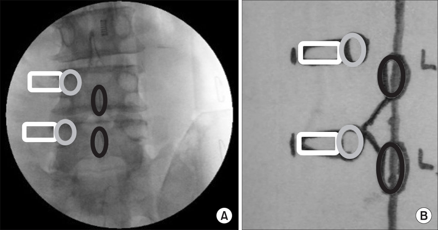
Figure 2. The anatomical lanmarks on true anteroposterior view of image intensifier during extraforaminal approach. (A) Transverse process (white rectangle), pedicle (grey), and spinous process (black). (B) The actual drawing on the patient.

Figure 3. (A) Shoulder type lumbar intervertebral disc herniation. (B) Axillary type lumbar intervertebral disc herniation (black circle: herniated disc, black star: nerve root).
Reference
-
References
1. Deyo RA, Mirza SK. Clinical practice. Herniated lumbar intervertebral disk. N Engl J Med. 2016. 374:1763–72.2. Kapetanakis S, Gkasdaris G, Thomaidis T, Charitoudis G, Kazakos K. Comparison of quality of life between men and women who underwent Transforaminal Percutaneous Endoscopic Discectomy for lumbar disc herniation. Int J Spine Surg. 2017. 11:28.
Article3. Cummins J, Lurie JD, Tosteson TD. . Descriptive epidemiology and prior healthcare utilization of patients in the Spine Patient Outcomes Research Trial’s (SPORT) three observational cohorts: disc herniation, spinal stenosis, and degenerative spondylolisthesis. Spine (Phila Pa 1976). 2006. 31:806–14.4. Frymoyer JW. Lumbar disk disease: epidemiology. Instr Course Lect. 1992. 41:217–23.5. Ala-Kokko L. Genetic risk factors for lumbar disc disease. Ann Med. 2002. 34:42–7.
Article6. Delgado-López PD, Rodríguez-Salazar A, Martín-Alonso J, Martín-Velasco V. [Lumbar disc herniation: natural history, role of physical examination, timing of surgery, treatment options and conflicts of interests]. Neurocirugia (Astur). 2017. 28:124–34. Spanish..
Article7. Kambin P, Zhou L. History and current status of percutaneous arthroscopic disc surgery. Spine (Phila Pa 1976). 1996. 21:57S–61S.8. You JW. [Lumbar disc disease]. J Korean Soc Spine Surg. 1999. 6:208–19. Korean..9. Owens RK 2nd, Carreon LY, Bisson EF, Bydon M, Potts EA, Glassman SD. Back pain improves significantly following discectomy for treatment of lumbar disc herniation. Spine J. 2017. 17:S68.
Article10. Caspar W. A new surgical procedure for lumbar disc herniation causing less tissue damage through a microsurgical approach. Wüllenweber R, Brock M, Hamer J, editors. Lumbar disc. Adult hydrocephalus. Heidelberg;Springer: 1977. 74–80.
Article11. Goald HJ. Microlumbar discectomy: followup of 147 patients. Spine (Phila Pa 1976). 1978. 3:183–5.12. Clark AJ, Safaee MM, Khan NR, Brown MT, Foley KT. Tubular microdiscectomy: techniques, complication avoidance, and review of the literature. Neurosurg Focus. 2017. 43:E7.
Article13. Kambin P. Percutaneous lumbar diskectomy. JAMA. 1989. 262:1776.
Article14. Mixter WJ, Barr JS. Rupture of the intervertebral disc with involvement of the spinal canal. J Neurosurg. 1964. 21:74–81.
Article15. Dandy WE. Loose cartilage from intervertebral disk simulating tumor of the spinal cord. Clin Orthop Relat Res. 1989. 238:4–8.
Article16. Maroon JC, Abla A, Bost J. Association between peridural scar and persistent low back pain after lumbar discectomy. Neurol Res. 1999. 21(Suppl 1):S43–6.
Article17. Williams RW. Microlumbar discectomy: a conservative surgical approach to the virgin herniated lumbar disc. Spine (Phila Pa 1976). 1978. 3:175–82.18. Casimiro M. Short-term outcome comparison between full-endoscopic interlaminar approach and open minimally invasive microsurgical technique for treatment of lumbar disc herniation. World Neurosurg. 2017. 108:894–900.e1.
Article19. Hu ZJ, Fang XQ, Zhou ZJ, Wang JY, Zhao FD, Fan SW. Effect and possible mechanism of muscle-splitting approach on multifidus muscle injury and atrophy after posterior lumbar spine surgery. J Bone Joint Surg Am. 2013. 95:e192.
Article20. Kambin P, Brager MD. Percutaneous posterolateral discectomy. Anatomy and mechanism. Clin Orthop Relat Res. 1987. 223:145–54.
Article21. Ahn SS, Kim SH, Kim DW, Lee BH. Comparison of outcomes of percutaneous endoscopic lumbar discectomy and open lumbar microdiscectomy for young adults: a retrospective matched cohort study. World Neurosurg. 2016. 86:250–8.
Article22. Choi KC, Shim HK, Park CJ, Lee DC, Park CK. Usefulness of percutaneous endoscopic lumbar foraminoplasty for lumbar disc herniation. World Neurosurg. 2017. 106:484–92.
Article23. Schroeder HW, Nehlsen M. Value of high-definition imaging in neuroendoscopy. Neurosurg Rev. 2009. 32:303–8.
Article24. Kim HS, Park JY. Comparative assessment of different percutaneous endoscopic interlaminar lumbar discectomy (PEID) techniques. Pain Physician. 2013. 16:359–67.25. Foley KT, Smith MM. Microendoscopic discectomy. Tech Neurosurg. 1997. 3:301–7.26. Choi DJ, Choi CM, Jung JT, Lee SJ, Kim YS. Learning curve associated with complications in biportal endoscopic spinal surgery: challenges and strategies. Asian Spine J. 2016. 10:624–9.
Article27. Teli M, Lovi A, Brayda-Bruno M. . Higher risk of dural tears and recurrent herniation with lumbar micro-endoscopic discectomy. Eur Spine J. 2010. 19:443–50.
Article28. Eun SS, Eum JH, Lee SH, Sabal LA. Biportal endoscopic lumbar decompression for lumbar disk herniation and spinal canal stenosis: a technical note. J Neurol Surg A Cent Eur Neurosurg. 2017. 78:390–6.29. Kambin P, Zhou L. Arthroscopic discectomy of the lumbar spine. Clin Orthop Relat Res. 1997. 337:49–57.
Article30. Perez-Cruet MJ, Foley KT, Isaacs RE. . Microendo-scopic lumbar discectomy: technical note. Neurosurgery. 2002. 51:S129–36.
Article31. Sairyo K, Sakai T, Higashino K, Inoue M, Yasui N, Dezawa A. Complications of endoscopic lumbar decompression surgery. Minim Invasive Neurosurg. 2010. 53:175–8.
Article32. Soliman HM. Irrigation endoscopic discectomy: a novel percutaneous approach for lumbar disc prolapse. Eur Spine J. 2013. 22:1037–44.
Article33. Choi DJ, Jung JT, Lee SJ, Kim YS, Jang HJ, Yoo B. Biportal endoscopic spinal surgery for recurrent lumbar disc herniations. Clin Orthop Surg. 2016. 8:325–9.
Article34. Hwa Eum J, Hwa Heo D, Son SK, Park CK. Percutaneous bi-portal endoscopic decompression for lumbar spinal stenosis: a technical note and preliminary clinical results. J Neurosurg Spine. 2016. 24:602–7.35. Joswig H, Richter H, Haile SR, Hildebrandt G, Fournier JY. Introducing interlaminar full-endoscopic lumbar diskectomy: a critical analysis of complications, recurrence rates, and outcome in view of two spinal surgeons’ learning curves. J Neurol Surg A Cent Eur Neurosurg. 2016. 77:406–15.36. Ahn JS, Lee HJ, Choi DJ, Lee KY, Hwang SJ. Extraforaminal approach of biportal endoscopic spinal surgery: a new endoscopic technique for transforaminal decompression and discectomy. J Neurosurg Spine. 2018. 28:492–8.
Article37. Soliman HM. Irrigation endoscopic decompressive laminotomy. A new endoscopic approach for spinal stenosis decompression. Spine J. 2015. 15:2282–9.38. Sencer A, Yorukoglu AG, Akcakaya MO. . Fully endoscopic interlaminar and transforaminal lumbar discectomy: short-term clinical results of 163 surgically treated patients. World Neurosurg. 2014. 82:884–90.
Article39. Young PM, Fenton DS, Czervionke LF. Postoperative annular pseudocyst: report of two cases with an unusual complication after microdiscectomy, and successful treatment by percutaneous aspiration and steroid injection. Spine J. 2009. 9:e9–15.
Article40. Kang SH, Park SW. Symptomatic post-discectomy pseudocyst after endoscopic lumbar discectomy. J Korean Neurosurg Soc. 2011. 49:31–6.
Article41. Ahn Y. Transforaminal percutaneous endoscopic lumbar discectomy: technical tips to prevent complications. Expert Rev Med Devices. 2012. 9:361–6.
Article
- Full Text Links
-
- Actions
-
Cited
- CITED
-
- Close
- Share
-
- Similar articles
-
- Complications and Management of Endoscopic Spinal Surgery
- Paramedian Unilateral ‘Bitubular’ Endoscopic Access for a Far Lateral Disc Herniation: A Novel Approach for Far Lateral Lumbar Pathologies
- Unilateral Biportal Endoscopic Translaminar Keyhole Approach to Treat High-grade Up-migrated Lumbar Disc Herniation: Technical Note
- Spinal Nerve Root Swelling Mimicking Intervertebral Disc Herniation in Magnetic Resonance Imaging: A Case Report
- Current Status of Biportal Endoscopic Decompression for Lumbar Central Stenosis
