Fibroma of the Extensor Digitorum Longus and Extensor Digitorum Brevis Conjoined Tendon Sheath: A Case Report
- Affiliations
-
- 1Department of Orthopaedic Surgery, Kangbuk Samsung Hospital, Sungkyunkwan University School of Medicine, Seoul, Korea. qortn97@naver.com
- 2Department of Orthopaedic Surgery, Konkuk University Chungju Hospital, Konkuk University College of Medicine, Chungju, Korea.
- KMID: 2449676
- DOI: http://doi.org/10.14193/jkfas.2019.23.2.74
Abstract
- Fibroma of the tendon sheath (FTS) was initially described in 1936 by Geschickter and Copeland as a benign firmed soft tissue tumor that is rare and less common than another soft tissue tumors, especially giant cell tumors (GCT) of the tendon sheath. The common distinct feature is a slow-growing least painful rare entity arising from the tendon or tendon sheath. FTS is detected mostly in the fingers, hands and wrists but less commonly in the foot. Very few cases of FTS have been described arising from a flexor tendon of the foot. This article describes a 51-year-old patient with FTS that developed in the extensor tendon of the foot, which is the only known FTS to form in this area. Heterogeneous low signal intensity in both the T1- and T2-weighted images was observed in magnetic resonance imaging. The lesion was excised completely by open surgery. Histologically, it showed randomly arranged, fibroblast-like spindle cells in dense fibrous tissue and had insufficient hemosiderin-laden macrophages that are typical for GCT.
MeSH Terms
Figure
-
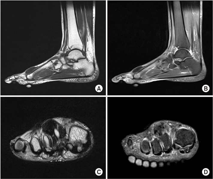
Figure 1 Magnetic resonance imaging (MRI) findings. (A) The tumor was well-defined heterogeneous low signal intensity on sagittal T1-weighted MRI image, located in the dorsal side of 2nd metatarsophalangeal (MTP) joint adjacent to the extensor digitorum longus (EDL) and extensor digitorum brevis (EDB). (B) The tumor shows same heterogeneous low signal intensity on sagittal T2-weighted MRI image. (C) Coronal T1-weighted MRI image revealing a mass firmly adherent to the dorsal side of 2nd MTP joint capsule and conjoined tendon of EDL and EDB. (D) The tumor on coronal T2-weighted MRI image shows same heterogeneous low signal intensity.

Figure 2 Gross photo. (A) Subcutaneous nodule protruded at dorsal side of 2nd metatarsophalangeal joint when patient flexion his toes. (B) At section, the mass appears to be white-tan, multilobular, and rubbery.
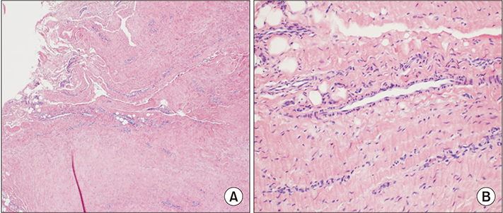
Figure 3 (A) Histologic image at low power showing a hypocellular mass of spindle cells within abundant collagenous stroma (H&E stain, ×40). (B) Histologic image at high power showing spindle cells with elongated nuclei, fine chromatin, small nucleoli, and eosinophilic collagenous stroma (H&E stain, ×100).
Reference
-
1. Geschickter CF, Copeland MM. Tumors of bone. New York: The American Journal of Cancer;1931.2. Chung EB, Enzinger FM. Fibroma of tendon sheath. Cancer. 1979; 44:1945–1954.
Article3. Kirby EJ, Shereff MJ, Lewis MM. Soft-tissue tumors and tumorlike lesions of the foot. An analysis of eighty-three cases. J Bone Joint Surg Am. 1989; 71:621–621.
Article4. Lu H, Chen Q, Shen H, Shen XQ, Wu SC. Fibroma of tendon sheath in planta. Springerplus. 2016; 5:575. DOI: 10.1186/s40064-016-2260-z.
Article5. Hintermann B, Nigg BM, Sommer C. Foot movement and tendon excursion: an in vitro study. Foot Ankle Int. 1994; 15:386–395. DOI: 10.1177/107110079401500708.
Article6. Nason GJ, Baker JF, O'Toole P, Walsh A, Lui DF, O'Neill F, et al. Fibroma of the peroneus longus tendon sheath in a child: a case report. J Orthop Surg (Hong Kong). 2013; 21:387–390. DOI: 10.1177/230949901302100326.
Article7. Fox MG, Kransdorf MJ, Bancroft LW, Peterson JJ, Flemming DJ. MR imaging of fibroma of the tendon sheath. AJR Am J Roentgenol. 2003; 180:1449–1453. DOI: 10.2214/ajr.180.5.1801449.
Article8. Humphreys S, McKee PH, Fletcher CD. Fibroma of tendon sheath: a clinicopathologic study. J Cutan Pathol. 1986; 13:331–338. DOI: 10.1111/j.1600-0560.1986.tb00467.x.
Article9. Ciatti R, Mariani PP. Fibroma of tendon sheath located within the ankle joint capsule. J Orthop Traumatol. 2009; 10:147–150. DOI: 10.1007/s10195-009-0058-2.
Article10. Hashimoto H, Tsuneyoshi M, Daimaru Y, Ushijima M, Enjoji M. Fibroma of tendon sheath: a tumor of myofibroblasts. A clinicopathologic study of 18 cases. Acta Pathol Jpn. 1985; 35:1099–1107.
- Full Text Links
-
- Actions
-
Cited
- CITED
-
- Close
- Share
-
- Similar articles
-
- Muscular Variations of Extensor Digitorum Brevis Muscle Related with Anterior Tarsal Tunnel Syndrome
- Split Transfer of Extensor Digitorum Longus of the Second Toe for Elongation of EHB after Modified Jones' Procedure
- Extensor Indicis Brevis: A Case Report
- An Extensor Digitorum Muscle for Index Finger Originated from the Extensor Carpi Radialis Brevis
- Extensor Pollicis Longus Tendon Rupture with Concomitant Rupture of the Extensor Digitorum Communis II Tendon and Extensor Indicis Proprius after Volar Plating for Distal Radius Fracture
