Neurointervention.
2019 Mar;14(1):9-16. 10.5469/neuroint.2018.01095.
Embryological Consideration of Dural AVFs in Relation to the Neural Crest and the Mesoderm
- Affiliations
-
- 1Department of Neurosurgery, Kameda Medical Center, Kamogawa, Japan. michihiro.tanaka@gmail.com
- KMID: 2446777
- DOI: http://doi.org/10.5469/neuroint.2018.01095
Abstract
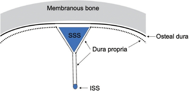
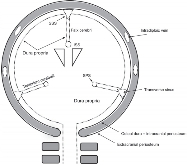
- Intracranial and spinal dural arteriovenous fistulas (DAVFs) are vascular pathologies of the dural membrane with arteriovenous shunts. They are abnormal communications between arteries and veins or dural venous sinuses that sit between the two sheets of the dura mater. The dura propria faces the surface of brain, and the osteal dura faces the bone. The location of the shunt points is not distributed homogeneously on the surface of the dural membrane, but there are certain areas susceptible to DAVFs. The dura mater of the olfactory groove, falx cerebri, inferior sagittal sinus, tentorium cerebelli, and falx cerebelli, and the dura mater at the level of the spinal cord are composed only of dura propria, and these areas are derived from neural crest cells. The dura mater of the cavernous sinus, transverse sinus, sigmoid sinus, and anterior condylar confluence surrounding the hypoglossal canal are composed of both dura propria and osteal dura; this group is derived from mesoderm. Although the cause of this heterogeneity has not yet been determined, there are some specific characteristics and tendencies in terms of the embryological features. The possible reasons for the segmental susceptibility to DAVFs are summarized based on the embryology of the dura mater.
MeSH Terms
Figure
Cited by 1 articles
-
Where Did the Dura Mater Come from?
Dae Chul Suh
Neurointervention. 2020;15(1):2-3. doi: 10.5469/neuroint.2020.00045.
Reference
-
1. Lasjaunias P, Chiu M, Ter Brugge K, Tolia A, Hurth M, Bernstein M. Neurological manifestations of intracranial dural arteriovenous malformations. J Neurosurg. 1986; 64:724–730.
Article2. Miller TR, Gandhi D. Intracranial dural arteriovenous fistulae: clinical presentation and management strategies. Stroke. 2015; 46:2017–2025.
Article3. Söderman M, Pavic L, Edner G, Holmin S, Andersson T. Natural history of dural arteriovenous shunts. Stroke. 2008; 39:1735–1739.
Article4. Baltsavias G, Valavanis A. Endovascular treatment of 170 consecutive cranial dural arteriovenous fistulae: results and complications. Neurosurg Rev. 2014; 37:63–71.
Article5. Zhao J, Xu F, Ren J, Manjila S, Bambakidis NC. Dural arteriovenous fistulas at the craniocervical junction: a systematic review. J Neurointerv Surg. 2016; 8:648–653.
Article6. Gross BA, Du R. The natural history of cerebral dural arteriovenous fistulae. Neurosurgery. 2012; 71:594–602. ; discussion 602-603.
Article7. Gandhi D, Chen J, Pearl M, Huang J, Gemmete JJ, Kathuria S. Intracranial dural arteriovenous fistulas: classification, imaging findings, and treatment. AJNR Am J Neuroradiol. 2012; 33:1007–1013.
Article8. Tanaka M. Embryological consideration of dural AVF. Acta Neurochir Suppl. 2016; 123:169–176.
Article9. Geibprasert S, Pereira V, Krings T, Jiarakongmun P, Toulgoat F, Pongpech S, et al. Dural arteriovenous shunts: a new classification of craniospinal epidural venous anatomical bases and clinical correlations. Stroke. 2008; 39:2783–2794.10. Gupta A, Periakaruppan A. Intracranial dural arteriovenous fistulas: a review. Indian J Radiol Imaging. 2009; 19:43–48.
Article11. MacK J, Squier W, Eastman JT. Anatomy and development of the meninges: implications for subdural collections and CSF circulation. Pediatr Radiol. 2009; 39:200–210.
Article12. Adeeb N, Mortazavi MM, Tubbs RS, Cohen-Gadol AA. The cranial dura mater: a review of its history, embryology, and anatomy. Childs Nerv Syst. 2012; 28:827–837.
Article13. Aurboonyawat T, Pereira V, Krings T, Toulgoat F, Chiewvit P, Lasjaunias P. Patterns of the cranial venous system from the comparative anatomy invertebrates. Part III. The ventricular system and comparative anatomy of the venous outlet of spinal cord and its homology with the five brain vesicles. Interv Neuroradiol. 2008; 14:125–136.14. O’Rahilly R, Müller F. The development of the neural crest in the human. J Anat. 2007; 211:335–351.15. Morriss-Kay G, Ruberte E, Fukiishi Y. Mammalian neural crest and neural crest derivatives. Ann Anat. 1993; 175:501–507.
Article16. Osumi-Yamashita N, Eto K. Mammalian Cranial Neural Crest Cells and Facial Development: (cranial neural crest cells/facial development/mammal/retinoic acid receptor/gene expression). Develop Growth & Differ. 1990; 32:451–459.
Article17. O’Rahilly R, Müller F. The meninges in human development. J Neuropathol Exp Neurol. 1986; 45:588–608.18. Tubbs RS, Loukas M, Louis RG Jr, Shoja MM, Acakpo-Satchivi L, Blount JP, et al. Anatomy of the falcine venous plexus. J Neurosurg. 2007; 107:155–157.
Article19. Wiltse LL. Anatomy of the extradural compartments of the lumbar spinal canal. Peridural membrane and circumneural sheath. Radiol Clin North Am. 2000; 38:1177–1206.20. Nicholas DS, Weller RO. The fine anatomy of the human spinal meninges. A light and scanning electron microscopy study. J Neurosurg. 1988; 69:276–282.21. Lang J. Anterior cranial fossa, the approach to the orbit and the ethmoid bone. In : . Berlin: Springer Berlin Heidelberg;1981. p. 67–93.22. Jiang X, Iseki S, Maxson RE, Sucov HM, Morriss-Kay GM. Tissue origins and interactions in the mammalian skull vault. Dev Biol. 2002; 241:106–116.
Article23. Berenstein A, Lasjaunias P, ter Brugge KG. Dural Arteriovenous Shunts. Surgical Neuroangiography. Berlin: Springer Berlin Heidelberg;2004. p. 565–607.24. Spittau B, Millán DS, El-Sherifi S, Hader C, Singh TP, Motschall E, et al. Dural arteriovenous fistulas of the hypoglossal canal: systematic review on imaging anatomy, clinical findings, and endovascular management. J Neurosurg. 2015; 122:883–903.
Article25. Mizutani K, Akiyama T, Minami Y, Toda M, Fujiwara H, Jinzaki M, et al. Intraosseous venous structures adjacent to the jugular tubercle associated with an anterior condylar dural arteriovenous fistula. Neuroradiology. 2018; 60:487–496.
Article26. Le Douarin NM, Dupin E. The Neural Crest, a Fourth Germ Layer of the Vertebrate Embryo. Trainor PABT-NCC. Neural Crest Cells. Boston: Elsevier;2014. p. 3–26.27. Calloni GW, Le Douarin NM, Dupin E. High frequency of cephalic neural crest cells shows coexistence of neurogenic, melanogenic, and osteogenic differentiation capacities. Proc Natl Acad Sci U S A. 2009; 106:8947–8952.
Article28. Hirasawa T, Kuratani S. Evolution of the vertebrate skeleton: morphology, embryology, and development. Zoological Lett. 2015; 1:2.
Article29. Noden DM, Schneider RA. Neural crest cells and the community of plan for craniofacial development: historical debates and current perspectives. Adv Exp Med Biol. 2006; 589:1–23.30. Gagan JR, Tholpady SS, Ogle RC. Cellular dynamics and tissue interactions of the dura mater during head development. Birth Defects Res C Embryo Today. 2007; 81:297–304.
Article31. Wilde JJ, Petersen JR, Niswander L. Genetic, epigenetic, and environmental contributions to neural tube closure. Annu Rev Genet. 2014; 48:583–611.
Article32. Katoh H, Shibata S, Fukuda K, Sato M, Satoh E, Nagoshi N, et al. The dual origin of the peripheral olfactory system: placode and neural crest. Mol Brain. 2011; 4:34.
Article33. Quackenbush KE, Luna-Fineman S, Magee JF, Gundogan M, Golobi M, Irie T, et al. Neuroblastoma involvement of the falx cerebri. Pediatr Blood Cancer. 2009; 53:1337–1339.
Article34. Doro D, Liu A, Grigoriadis AE, Liu KJ. The osteogenic potential of the neural crest lineage may contribute to craniosynostosis. Mol Syndromol. 2019; 10:48–57.
Article35. Rodionov AA, Asfandiyarov RI. Morphogenesis of the epidural space in humans during the embryonic and early fetal periods. Neurosci Behav Physiol. 2010; 40:137–142.
Article36. Djindjian R, Merland JJ. Super-Selective Arteriography of the External Carotid Artery. Berlin:: Springer Berlin Heidelberg;1978.37. Lalwani AK, Dowd CF, Halbach V V. Grading venous restrictive disease in patients with dural arteriovenous fistulas of the transverse/sigmoid sinus. J Neurosurg. 1993; 79:11–15.
Article38. Cognard C, Gobin YP, Pierot L, Bailly AL, Houdart E, Casasco A, et al. Cerebral dural arteriovenous fistulas: clinical and angiographic correlation with a revised classification of venous drainage. Radiology. 1995; 194:671–680.
Article39. Gomez J, Amin AG, Gregg L, Gailloud P. Classification schemes of cranial dural arteriovenous fistulas. Neurosurg Clin N Am. 2012; 23:55–62.
Article40. Borden JA, Wu JK, Shucart WA. A proposed classification for spinal and cranial dural arteriovenous fistulous malformations and implications for treatment. J Neurosurg. 1995; 82:166–179.
Article41. Baltsavias G, Roth P, Valavanis A. Cranial dural arteriovenous shunts. Part 3. Classification based on the leptomeningeal venous drainage. Neurosurg Rev. 2015; 38:273–281.
Article42. Oyama Y, Kazama JJ, Fukagawa M, Arakawa Y, Ezuka I. Ectopic ossification in the cranial dura mater in dialysis patients with secondary hyperparathyroidism. NDT Plus. 2010; 3:64–67.
Article43. Kuratani S. Craniofacial development and the evolution of the vertebrates: the old problems on a new background. Zoolog Sci. 2005; 22:1–19.
Article44. Noden DM, Trainor PA. Relations and interactions between cranial mesoderm and neural crest populations. J Anat. 2005; 207:575–601.
Article45. Lekkhong E, Pongpech S, Ter Brugge K, Jiarakongmun P, Willinsky R, Geibprasert S, et al. Transvenous embolization of intracranial dural arteriovenous shunts through occluded venous segments: experience in 51 patients. AJNR Am J Neuroradiol. 2011; 32:1738–1744.
Article
- Full Text Links
- Actions
-
Cited
- CITED
-
- Close
- Share
- Similar articles
-
- 3 Cases of Neurofibromatosis Associated with Intracranial and Spinal Lesions
- Sox9 regulates development of neural crest and otic placode in a time- and dose-dependent fashion
- Immunohistochemistry: sole tool in diagnosing a rare case of primary vaginal amelanotic melanoma
- Understanding of Human Embryo Development for Teratogen Counselling
- Usefulness of sectional images in dural AVF for the interpretation of venous anatomy


