Usefulness of sectional images in dural AVF for the interpretation of venous anatomy
- Affiliations
-
- 1Department of Radiology, Dong-A University Hospital, Busan, Korea
- KMID: 2556974
- DOI: http://doi.org/10.7461/jcen.2023.E2022.10.001
Abstract
- Knowledge of the venous anatomy is essential for appropriately treating dural arteriovenous fistulas (AVFs). It is challenging to determine the overall venous structure despite performing selective angiography for dural AVFs with feeder from multiple selected arteries. This is because only a part of the veins can be observed through the shunt in the selected artery. Therefore, after performing selective angiography of all vessels to understand the approximate venous anatomy, the venous anatomy can be easily understood by closely examining the source image of computed tomographic angiography or magnetic resonance angiography. Through this, it is possible to specify the vein that is to be blocked (target embolization), thereby avoiding extensive blocking of the vein and avoiding various complications. In the case of dural AVF with feeder from single selected artery, if the multiplanar reconstruction image of the three-dimensional rotational computed tomography obtained by performing angiography is analyzed thoroughly, a shunted pouch can be identified. If embolization is performed by targeting this area, unnecessary sinus total packing can be avoided.
Keyword
Figure
-

Fig. 1. Dural AVF in vein of Labbe. (A) ECA selective angiography, lateral view. (B) Microcatheter navigation into posterior branch of middle meningeal artery. (C) Transarterial ONYX embolization. (D) After treatment, total occlusion of dural AVF. AVF, arteriovenous fistula; ECA, external carotid artery
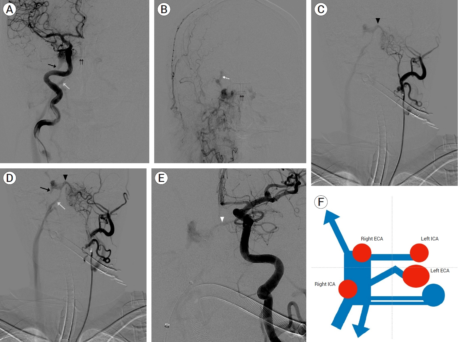
Fig. 2. Case 1. Dural AVF in right cavernous sinus. (A) Right ICA selective angiography, AP view. (B) Right ECA selective angiography, AP view. (C, D) Left ECA selective angiography, AP view. (E) Left ICA selective angiography, AP view. Non shunted venous pouch (black arrow). Right inferior petrosal sinus (white arrow). Intercavernous sinus (black double arrows). Right superior ophthalmic vein (white dot arrow). Shunted pouch connecting to left ECA (black arrow head). Shunted pouch connecting to left ICA (white arrow head) in A, B, C, D, and E. (F) Illustration of venous anatomy in dural AVF. ICA, internal carotid artery; AP, anteroposterior; ECA, external carotid artery; AVF, arteriovenous fistula
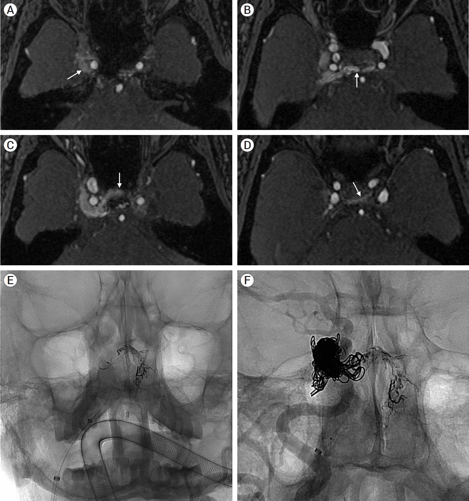
Fig. 3. Case 1. Source images of MRA in A, B, C, and D. (A) Non shunted pouch (white arrow). (B) Shunted pouch connecting to left ECA (white arrow). (C) Intercavernous sinus (white arrow). (D) Shunted pouch connecting to left ICA (white arrow). (E, F) Transvenous ONYX and coil embolization of dural AVF in cavernous sinus. MRA, magnetic resonance angiography; ECA, external carotid artery; ICA, internal carotid artery; AVF, arteriovenous fistula
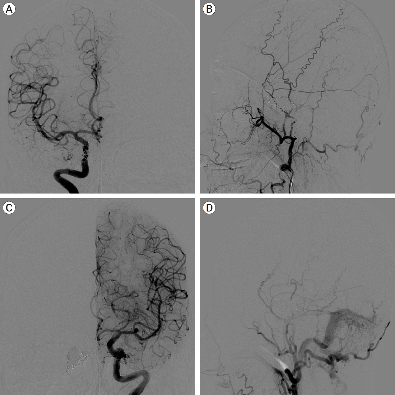
Fig. 4. Case 1. 1 year follow up angiography. No recurrence of dural AVF in right cavernous sinus. (A) Right ICA selective angiography, AP view. (B) Right ECA selective angiography, lateral view. (C) Left ICA selective angiography, AP view. Known aneurysm in left MCA bifurcation area which was coil-embolized. (D) Left ECA selective angiography, lateral view. De novo development of dural AVF in left transverse sinus. AVF, arteriovenous fistula; ICA, internal carotid artery; AP, anteroposterior; ECA, external carotid artery
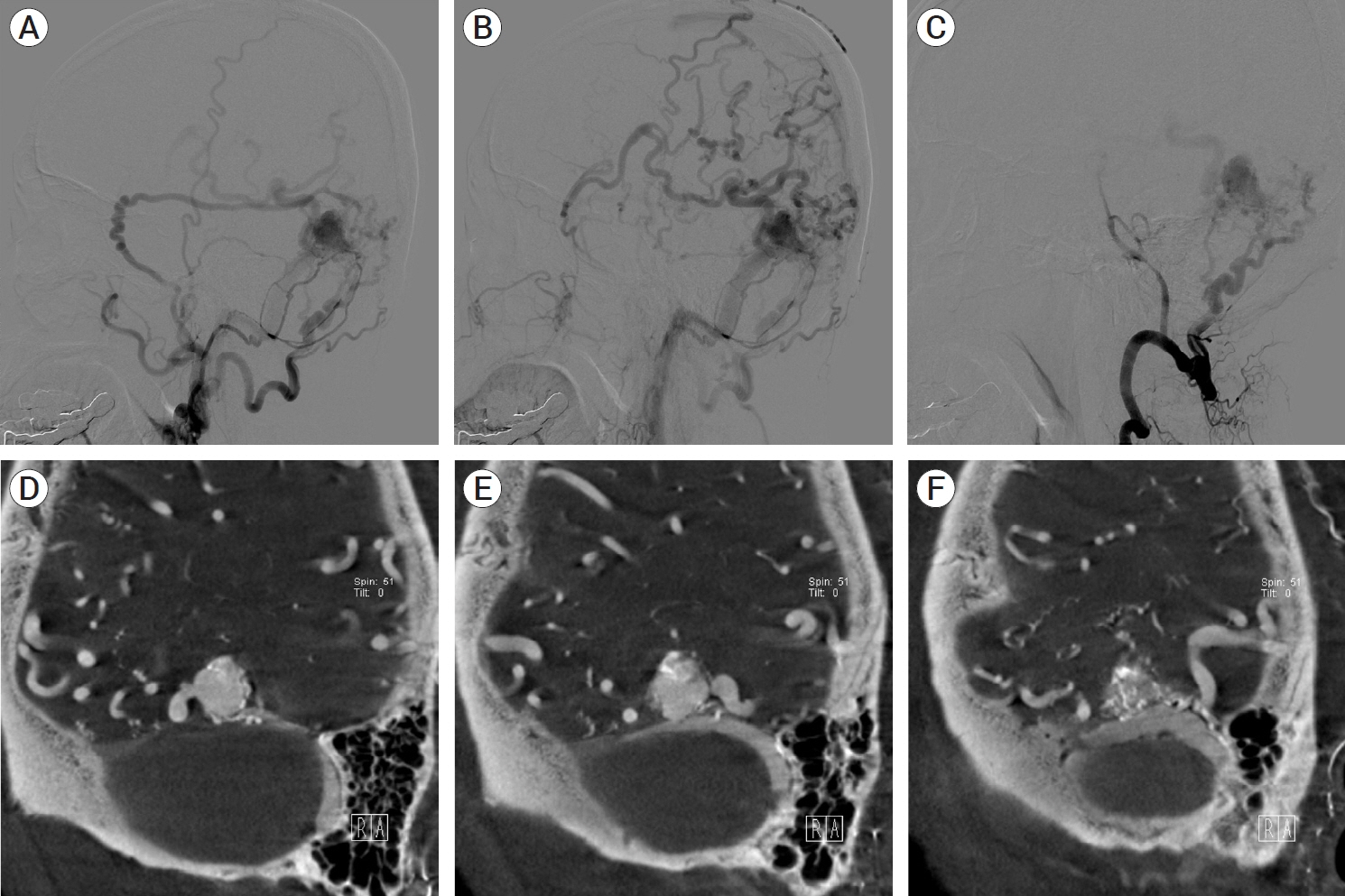
Fig. 5. Case 2. Dural AVF in left transeverse sinus. (A, B) Left ECA selective angiography, lateral view. (C) Left VA selective angiography, lateral view. (D, E, F) MPR images of 3D rotational CT. AVF, arteriovenous fistula; ECA, external carotid artery; VA, vertebral artery; MPR, multiplanar reconstruction; 3D, three-dimensional; CT, computed tomography
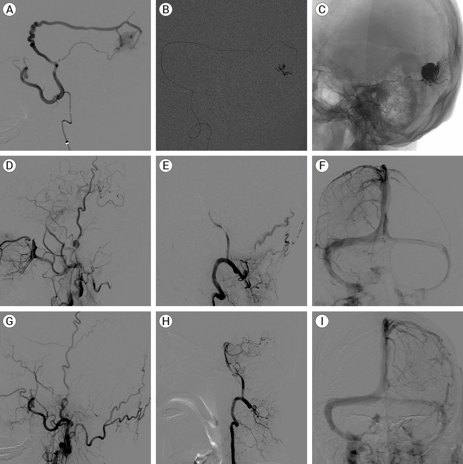
Fig. 6. Case 2. Transarteral ONYX embolization and follow up angiography. (A, B, C) Transarteral ONYX embolization of dural AVF in transeverse sinus. (D, E, F) After treatment, immediately follow up angiography. Total occlusion of dural AVF and preserved venous flow through left transeverse sinus. (G, H, I) 1 year follow up angiography. No recurrence of dural AVF and preserved venous flow through left transeverse sinus. AVF, arteriovenous fistula

Fig. 7. Case 3. Dural AVF in left transverse sinus and transvenous embolization. Left ECA selective angiography, lateral view in A and B. (A) Shunted pouch (black arrow). (B) Tight stenosis in junction of transverse and sigmoid sinus (black arrow). MPR images of 3D rotational CT in C, D, and E. (C) Occlusion in distal portion of transverse sinus (white arrow). (D) Shunted pouch (white arrow). (E) Tight stenosis in junction of transverse and sigmoid sinus (white arrow). (F) After passing through stenosis with microcatheter, stenosis was blocked by microcatheter. More detail of venous anatomy was visible. (G) Transvenous ONYX and coil embolization of dural AVF. (H) After treatment, immediately follow up angiography. Total occlusion of dural AVF. (I) 1 year follow up angiography, No recurrence of dural AVF. AVF, arteriovenous fistula; ECA, external carotid artery; MPR, multiplanar reconstruction; 3D, three-dimensional; CT, computed tomography
Reference
-
1. Bhatia KD, Lee H, Kortman H, Klostranec J, Guest W, Wälchli T, et al. Endovascular management of intracranial dural arteriovenous fistulas: Transarterial approach. AJNR Am J Neuroradiol. 2022; Mar. 43(3):324–31.
Article2. Bhatia KD, Lee H, Kortman H, Klostranec J, Guest W, Wälchli T, et al. Endovascular management of intracranial dural AVFs: Transvenous approach. AJNR Am J Neuroradiol. 2022; Apr. 43(4):510–6.
Article3. Kiyosue H, Tanoue S, Hori Y, Hongo N, Mori H. Shunted pouches of cavernous sinus dural AVFs: Evaluation by 3D rotational angiography. Neuroradiology. 2015; Mar. 57(3):283–90.
Article4. Kiyosue H, Tanoue S, Okahara M, Hori Y, Kashiwagi J, Sagara Y, et al. Angioarchitecture of transverse-sigmoid sinus dural arteriovenous fistulas: evaluation of shunted pouches by multiplanar reformatted images of rotational angiography. AJNR Am J Neuroradiol. 2013; Aug. 34(8):1612–20.
Article5. Tagawa M, Inoue A, Murayama K, Matsumoto S, Ozaki S, Nishikawa M, et al. Utility of targeted balloon protection of the venous sinus for endovascular treatment of dural arteriovenous fistula by transarterial embolization with Onyx: A case report and literature review. Surg Neurol Int. 2021; Jul. 12:340.
Article6. Torok CM, Nogueira RG, Yoo AJ, Leslie-Mazwi TM, Hirsch JA, Stapleton CJ, et al. Transarterial venous sinus occlusion of dural arteriovenous fistulas using ONYX. Interv Neuroradiol. 2016; Dec. 22(6):711–6.
Article
- Full Text Links
-
- Actions
-
Cited
- CITED
-
- Close
- Share
-
- Similar articles
-
- Iatrogenic mixed pial and dural arteriovenous fistula after pterional approach for surgical clipping of aneurysm: A case report
- Transvenous coil embolization of hypoglossal canal dural arteriovenous fistula using detachable coils: A case report
- A Rare Case of Subarachnoid Hemorrhage caused by Ruptured Venous Varix Due to Dural Arteriovenous Fistula at the Foramen Magnum Fed Solely by the Ascending Pharyngeal Artery
- Surgical Treatment of Carotid-Cavernous Fistula and Intracranial Dural Arteriovenous Malformations
- Delayed Dural Arteriovenous Fistula after Microvascular Decompression for Hemifacial Spasm
