Pediatr Gastroenterol Hepatol Nutr.
2019 May;22(3):298-302. 10.5223/pghn.2019.22.3.298.
Herpetic Esophagitis in Immunocompetent Child
- Affiliations
-
- 1Department of Pediatrics, Faculty of Medicine, Jordan University of Science and Technology, Irbid, Jordan. emaltamimi@just.edu.jo
- 2Department of Pathology, Faculty of Medicine, Jordan University of science and Technology, Irbid, Jordan.
- KMID: 2444856
- DOI: http://doi.org/10.5223/pghn.2019.22.3.298
Abstract
- A previously healthy 2.5-year-old male child presented with vomiting, diarrhea, and fever. During hospitalization he developed odynophagia and refusal to eat. His symptoms did not respond to acid suppressant therapy. He underwent upper endoscopy which showed severe inflammation, ulcerations and abundant necrosis. Histopathological features and serological testing were consistent with herpetic esophagitis. He had no history of recurrent infections or history of sick contacts. His immunological work up showed normal level of immunoglobulins and his White Blood Cells subpopulations were normal. His HSV serology was positive. The patient was started on acyclovir 5 mg/kg q 8 hours. He resolved his symptoms within 24 hours of treatment.
Keyword
MeSH Terms
Figure
-
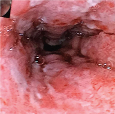
Fig. 1 Endoscopic examination reveals severe inflammation, with ulceration and exudate.

Fig. 2 Histopathologic examination of the esophageal mucosa biopsy (hematoxylin and eosin staining). (A) Fragments of ulcer slough and granulation tissue, are also observed. Few cells exhibit cytopathic changes suggestive of herpetic viral etiology; in the form of multinucleation, chromatin margination, and vague nuclear inclusions. Denuded mucosa, ulcer slough, and granulation can be observed (100×). (B) A high-power view (400×) of the same tissue fragment, shows viral cytopathic changes (multinucleation, chromatin margination, and vague nuclear inclusions).
Reference
-
1. Baehr PH, McDonald GB. Esophageal infections: risk factors, presentation, diagnosis, and treatment. Gastroenterology. 1994; 106:509–532.
Article2. Ramanathan J, Rammouni M, Baran J Jr, Khatib R. Herpes simplex virus esophagitis in the immunocompetent host: an overview. Am J Gastroenterol. 2000; 95:2171–2176.
Article3. Rodrigues F, Brandão N, Duque V, Ribeiro C, António AM. Herpes simplex virus esophagitis in immunocompetent children. J Pediatr Gastroenterol Nutr. 2004; 39:560–563.
Article4. Al-Hussaini AA, Fagih MA. Herpes simplex ulcerative esophagitis in healthy children. Saudi J Gastroenterol. 2011; 17:353–356.
Article5. Galbraith JC, Shafran SD. Herpes simplex esophagitis in the immunocompetent patient: report of four cases and review. Clin Infect Dis. 1992; 14:894–901.
Article6. Fritz J, Lerner D, Suchi M. Herpes simplex virus esophagitis in immunocompetent children: a harbinger of eosinophilic esophagitis? J Pediatr Gastroenterol Nutr. 2018; 66:609–613.
Article
- Full Text Links
-
- Actions
-
Cited
- CITED
-
- Close
- Share
-
- Similar articles
-
- A Case of Herpes simplex Esophagitis in an Immunocompetent Boy
- A Case of Herpes Esophagitis Confirmed by Electron Microscopic Findings
- Viral Infection in Upper Gastrointestinal Tract
- Herpes Simplex Esophagitis: A report of two cases
- A case of cytomegalovirus esophagitis associated with upper gastrointestinal bleeding
