J Korean Orthop Assoc.
2019 Apr;54(2):172-176. 10.4055/jkoa.2019.54.2.172.
Symptomatic Calcific Deposition in Posterior Cruciate Ligament of the Knee
- Affiliations
-
- 1Departments of Orthopedic Surgery, College of Medicine, Catholic Kwandong University, Incheon, Korea. sysung@ish.ac.kr
- KMID: 2444783
- DOI: http://doi.org/10.4055/jkoa.2019.54.2.172
Abstract
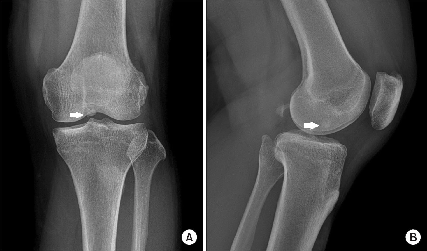
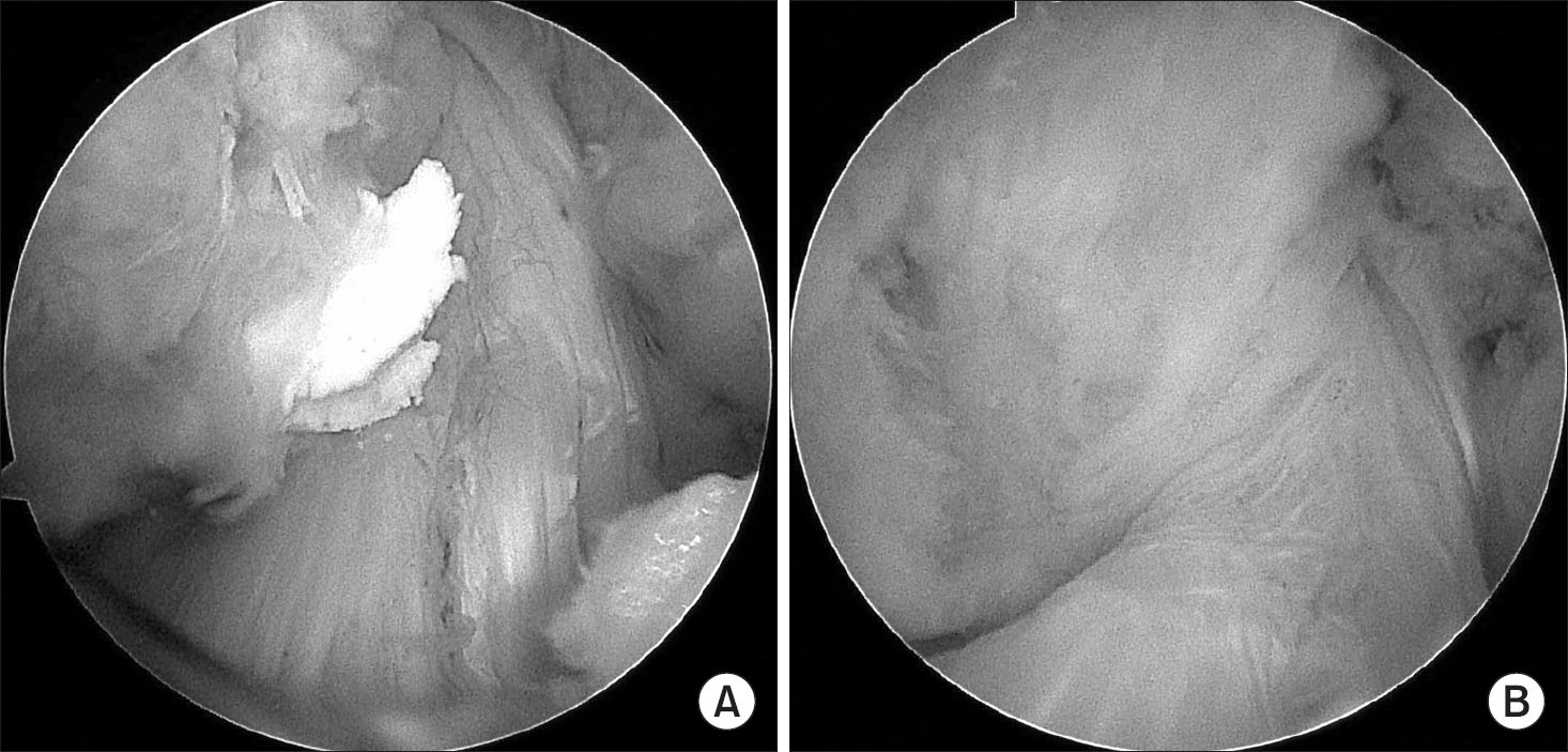
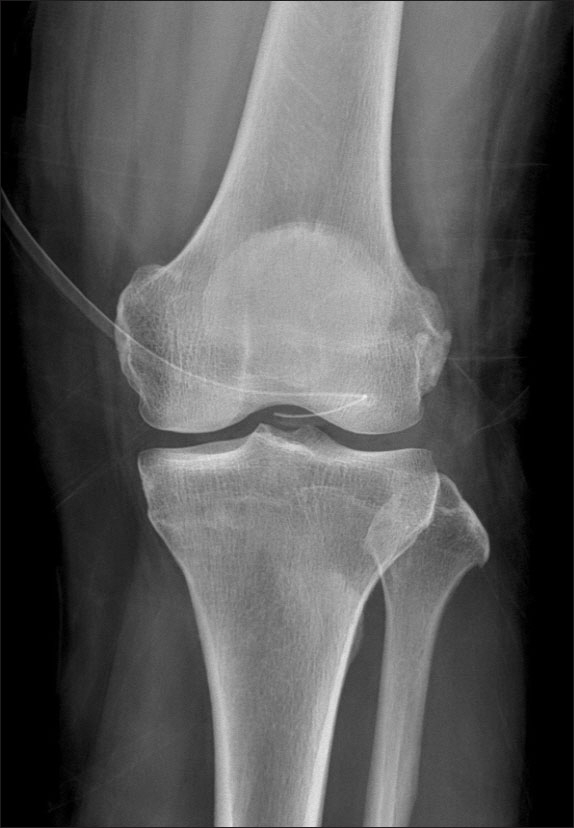
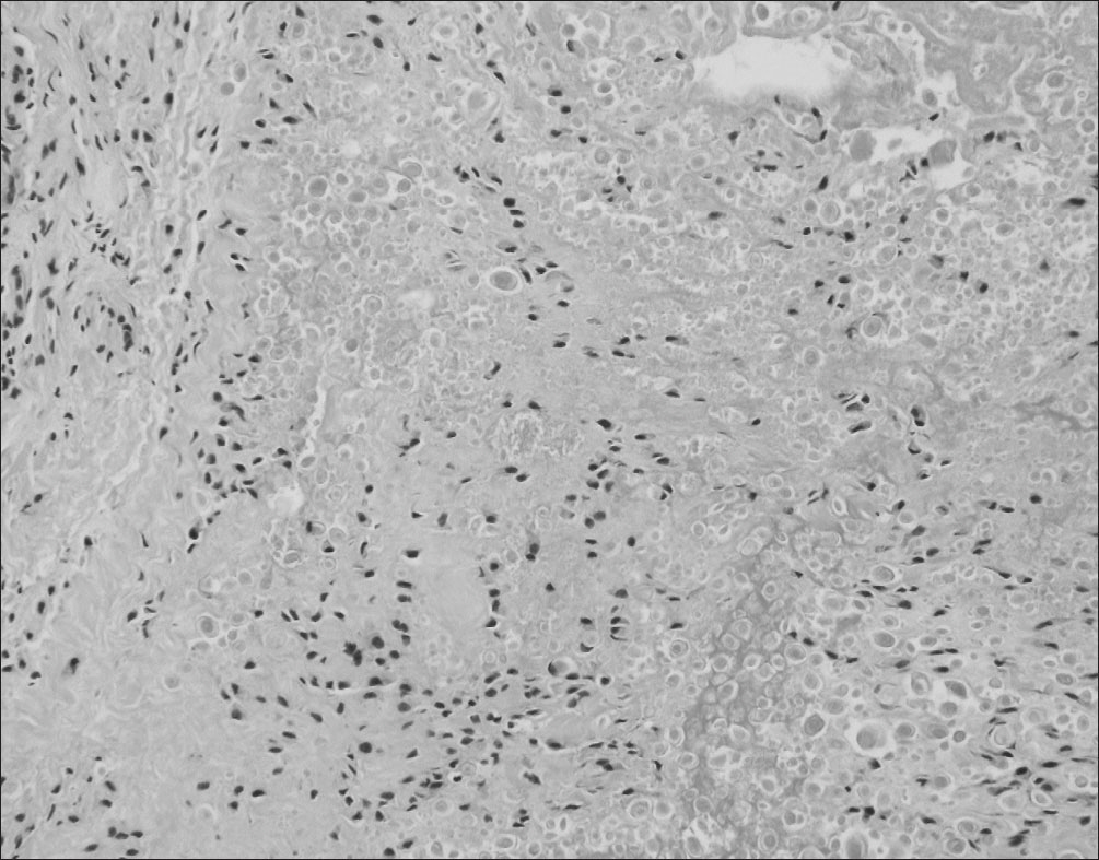
- Calcium deposition disease, including calcific tendinitis, rarely affects the knee joint. Only a few cases can be found in the literatures and there is no case report of symptomatic calcific deposition arising from the posterior cruciate ligament in Korea. The authors encountered a case of symptomatic calcific deposition arising from the posterior cruciate ligament, which was excised arthroscopically and confirmed pathologically. This paper reports this case with a review of the relevant literature.
Keyword
Figure
Reference
-
References
1. Chan R, Kim DH, Millett PJ, Weissman BN. Calcifying tendinitis of the rotator cuff with cortical bone erosion. Skeletal Radiol. 2004. 33:596–9.
Article2. Uhthoff HK, Loehr JW. Calcific tendinopathy of the rotator cuff: pathogenesis, diagnosis, and management. J Am Acad Orthop Surg. 1997. 5:183–91.
Article3. Holt PD, Keats TE. Calcific tendinitis: a review of the usual and unusual. Skeletal Radiol. 1993. 22:1–9.
Article4. McKendry RJ, Uhthoff HK, Sarkar K, Hyslop PS. Calcifying tendinitis of the shoulder: prognostic value of clinical, histologic, and radiologic features in 57 surgically treated cases. J Rheumatol. 1982. 9:75–80.5. Shenoy PM, Kim DH, Wang KH. . Calcific tendinitis of popliteus tendon: arthroscopic excision and biopsy. Orthopedics. 2009. 32:127.
Article6. Tibrewal SB. Acute calcific tendinitis of the popliteus tendon: an unusual site and clinical syndrome. Ann R Coll Surg Engl. 2002. 84:338–41.7. Chan W, Chase HE, Cahir JG, Walton NP. Calcific tendinitis of biceps femoris: an unusual site and cause for lateral knee pain. BMJ Case Rep. 2016. 2016. pii: bcr2016215745.
Article8. Song K, Dong J, Zhang Y. . Arthroscopic management of calcific tendonitis of the medial collateral ligament. Knee. 2013. 20:63–5.
Article9. Molloy ES, McCarthy GM. Hydroxyapatite deposition disease of the joint. Curr Rheumatol Rep. 2003. 5:215–21.
Article10. Kim MK, Bae JH, Jeon YS. Conservative and early ar-throscopic treatment of calcific tendinitis. J Korean Ar-throscop Soc. 2009. 13:149–54.
- Full Text Links
- Actions
-
Cited
- CITED
-
- Close
- Share
- Similar articles
-
- Ganglion Cyst of the Posterior Cruciate Ligament: A Case Report
- Symptomatic Posterior Cruciate Ganglion Cyst Causing Impingement between Posterior Root of the Medial Meniscus and Anterior to the Posterior Cruciate Ligament
- Reconstruction of the Posterior Cruciate Ligament Using the Medial Meniscus
- Extension Block by the Posterior Cruciate Ligament Partial Rupture in the Knee: A Case Report
- Symptomatic Mid-Substance Posterior Cruciate Ligament Calcification of the Knee Joint


