J Periodontal Implant Sci.
2019 Apr;49(2):90-104. 10.5051/jpis.2019.49.2.90.
Periodontal regenerative therapy in endo-periodontal lesions: a retrospective study over 5 years
- Affiliations
-
- 1Department of Conservative Dentistry, Kyung Hee University Dental Hospital, Seoul, Korea.
- 2Department of Dental Biomaterials Science and Dental Research Institute, Seoul National University School of Dentistry, Seoul, Korea.
- 3Department of Dentistry and Periodontology, Hanyang University College of Medicine, Seoul, Korea. hjyperio@hanyang.ac.kr
- KMID: 2444221
- DOI: http://doi.org/10.5051/jpis.2019.49.2.90
Abstract
- PURPOSE
The aim of this study was to evaluate clinical and radiographic changes and the survival rate after periodontal surgery using deproteinized bovine bone mineral (DBBM) with 10% collagen or DBBM with a collagen membrane in endo-periodontal lesions.
METHODS
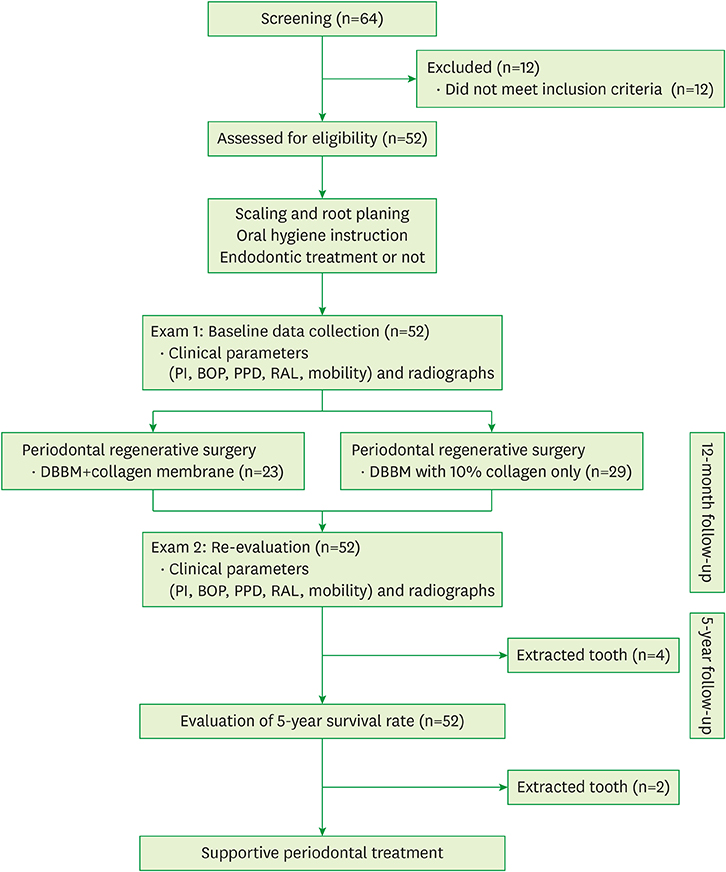
A total of 52 cases (41 patients) with at least 5 years of follow-up were included in this study. After scaling and root planing with or without endodontic treatment, periodontal regenerative procedures with DBBM with 10% collagen alone or DBBM with a collagen membrane were performed, yielding the DBBM + 10% collagen and DBBM + collagen membrane groups, respectively. Changes in clinical parameters including the plaque index, bleeding on probing, probing pocket depth, gingival recession, relative clinical attachment level, mobility, and radiographic bone gains were evaluated immediately before periodontal surgical procedures and at a 12-month follow-up.
RESULTS
At the 12-month follow-up after regenerative procedures, improvements in clinical parameters and radiographic bone gains were observed in both treatment groups. The DBBM + 10% collagen group showed greater probing pocket depth reduction (4.52±1.06 mm) than the DBBM + collagen membrane group (4.04±0.82 mm). However, there were no significant differences between the groups. Additionally, the radiographic bone gain in the DBBM + 10% collagen group (5.15±1.54 mm) was comparable to that of the DBBM + collagen membrane group (5.35±1.84 mm). The 5-year survival rate of the teeth with endo-periodontal lesions after periodontal regenerative procedures was 92.31%.
CONCLUSIONS
This study showed that regenerative procedures using DBBM with 10% collagen alone improved the clinical attachment level and radiographic bone level in endo-periodontal lesions. Successful maintenance of the results after regenerative procedures in endo-periodontal lesions can be obtained by repeated oral hygiene education within strict supportive periodontal treatment.
Keyword
MeSH Terms
Figure
Reference
-
1. Löe H, Theilade E, Jensen SB. Experimental gingivitis in man. J Periodontol. 1965; 36:177–187.
Article2. Zehnder M, Gold SI, Hasselgren G. Pathologic interactions in pulpal and periodontal tissues. J Clin Periodontol. 2002; 29:663–671.
Article3. Fabricius L, Dahlén G, Sundqvist G, Happonen RP, Möller AJ. Influence of residual bacteria on periapical tissue healing after chemomechanical treatment and root filling of experimentally infected monkey teeth. Eur J Oral Sci. 2006; 114:278–285.
Article4. Simon JH, Glick DH, Frank AL. The relationship of endodontic-periodontic lesions. J Periodontol. 1972; 43:202–208.
Article5. Herrera D, Retamal-Valdes B, Alonso B, Feres M. Acute periodontal lesions (periodontal abscesses and necrotizing periodontal diseases) and endo-periodontal lesions. J Periodontol. 2018; 89:Suppl 1. S85–S102.
Article6. Hirsch JM, Ahlström U, Henrikson PA, Heyden G, Peterson LE. Periapical surgery. Int J Oral Surg. 1979; 8:173–185.
Article7. Skoglund A, Persson G. A follow-up study of apicoectomized teeth with total loss of the buccal bone plate. Oral Surg Oral Med Oral Pathol. 1985; 59:78–81.
Article8. Kim E, Song JS, Jung IY, Lee SJ, Kim S. Prospective clinical study evaluating endodontic microsurgery outcomes for cases with lesions of endodontic origin compared with cases with lesions of combined periodontal-endodontic origin. J Endod. 2008; 34:546–551.
Article9. Kao RT, Nares S, Reynolds MA. Periodontal regeneration-intrabony defects: a systematic review from the AAP Regeneration Workshop. J Periodontol. 2015; 86:Suppl. S77–104.10. Cortellini P, Tonetti MS. Clinical concepts for regenerative therapy in intrabony defects. Periodontol 2000. 2015; 68:282–307.
Article11. Sculean A, Nikolidakis D, Nikou G, Ivanovic A, Chapple IL, Stavropoulos A. Biomaterials for promoting periodontal regeneration in human intrabony defects: a systematic review. Periodontol 2000. 2015; 68:182–216.
Article12. Nevins ML, Camelo M, Lynch SE, Schenk RK, Nevins M. Evaluation of periodontal regeneration following grafting intrabony defects with bio-oss collagen: a human histologic report. Int J Periodontics Restorative Dent. 2003; 23:9–17.13. Sculean A, Stavropoulos A, Windisch P, Keglevich T, Karring T, Gera I. Healing of human intrabony defects following regenerative periodontal therapy with a bovine-derived xenograft and guided tissue regeneration. Clin Oral Investig. 2004; 8:70–74.
Article14. Hartman GA, Arnold RM, Mills MP, Cochran DL, Mellonig JT. Clinical and histologic evaluation of anorganic bovine bone collagen with or without a collagen barrier. Int J Periodontics Restorative Dent. 2004; 24:127–135.15. Guillemin MR, Mellonig JT, Brunsvold MA. Healing in periodontal defects treated by decalcified freeze-dried bone allografts in combination with ePTFE membranes (I). Clinical and scanning electron microscope analysis. J Clin Periodontol. 1993; 20:528–536.
Article16. Brkovic BM, Prasad HS, Rohrer MD, Konandreas G, Agrogiannis G, Antunovic D, et al. Beta-tricalcium phosphate/type I collagen cones with or without a barrier membrane in human extraction socket healing: clinical, histologic, histomorphometric, and immunohistochemical evaluation. Clin Oral Investig. 2012; 16:581–590.
Article17. Lee JS, Jung JS, Im GI, Kim BS, Cho KS, Kim CS. Ridge regeneration of damaged extraction sockets using rhBMP-2: an experimental study in canine. J Clin Periodontol. 2015; 42:678–687.
Article18. Becker W, Becker BE. Clinical applications of guided tissue regeneration: surgical considerations. Periodontol 2000. 1993; 1:46–53.
Article19. Villar CC, Cochran DL. Regeneration of periodontal tissues: guided tissue regeneration. Dent Clin North Am. 2010; 54:73–92.
Article20. Anderegg CR, Metzler DG, Nicoll BK. Gingiva thickness in guided tissue regeneration and associated recession at facial furcation defects. J Periodontol. 1995; 66:397–402.
Article21. Silness J, Löe H. Periodontal disease in pregnancy. II. Correlation between oral hygiene and periodontal condition. Acta Odontol Scand. 1964; 22:121–135.
Article22. Cortellini P, Prato GP, Tonetti MS. The modified papilla preservation technique. A new surgical approach for interproximal regenerative procedures. J Periodontol. 1995; 66:261–266.
Article23. Cortellini P, Prato GP, Tonetti MS. The simplified papilla preservation flap. A novel surgical approach for the management of soft tissues in regenerative procedures. Int J Periodontics Restorative Dent. 1999; 19:589–599.24. Björn H, Halling A, Thyberg H. Radiographic assessment of marginal bone loss. Odontol Revy. 1969; 20:165–179.25. Liñares A, Cortellini P, Lang NP, Suvan J, Tonetti MS. European Research Group on Periodontology (ErgoPerio). Guided tissue regeneration/deproteinized bovine bone mineral or papilla preservation flaps alone for treatment of intrabony defects. II: radiographic predictors and outcomes. J Clin Periodontol. 2006; 33:351–358.
Article26. Tonetti MS, Pini Prato G, Williams RC, Cortellini P. Periodontal regeneration of human infrabony defects. III. Diagnostic strategies to detect bone gain. J Periodontol. 1993; 64:269–277.
Article27. Camelo M, Nevins ML, Schenk RK, Simion M, Rasperini G, Lynch SE, et al. Clinical, radiographic, and histologic evaluation of human periodontal defects treated with Bio-Oss and Bio-Gide. Int J Periodontics Restorative Dent. 1998; 18:321–331.28. Paolantonio M, Scarano A, Di Placido G, Tumini V, D'Archivio D, Piattelli A. Periodontal healing in humans using anorganic bovine bone and bovine peritoneum-derived collagen membrane: a clinical and histologic case report. Int J Periodontics Restorative Dent. 2001; 21:505–515.29. Camelo M, Nevins ML, Lynch SE, Schenk RK, Simion M, Nevins M. Periodontal regeneration with an autogenous bone-Bio-Oss composite graft and a Bio-Gide membrane. Int J Periodontics Restorative Dent. 2001; 21:109–119.30. Schmidt JC, Walter C, Amato M, Weiger R. Treatment of periodontal-endodontic lesions--a systematic review. J Clin Periodontol. 2014; 41:779–790.
Article31. Schulz A, Hilgers RD, Niedermeier W. The effect of splinting of teeth in combination with reconstructive periodontal surgery in humans. Clin Oral Investig. 2000; 4:98–105.
Article32. Cortellini P, Stalpers G, Mollo A, Tonetti MS. Periodontal regeneration versus extraction and prosthetic replacement of teeth severely compromised by attachment loss to the apex: 5-year results of an ongoing randomized clinical trial. J Clin Periodontol. 2011; 38:915–924.
Article33. Siciliano VI, Andreuccetti G, Siciliano AI, Blasi A, Sculean A, Salvi GE. Clinical outcomes after treatment of non-contained intrabony defects with enamel matrix derivative or guided tissue regeneration: a 12-month randomized controlled clinical trial. J Periodontol. 2011; 82:62–71.
Article34. Cortellini P, Paolo G, Prato P, Tonetti MS. Long-term stability of clinical attachment following guided tissue regeneration and conventional therapy. J Clin Periodontol. 1996; 23:106–111.
Article35. Eickholz P, Krigar DM, Kim TS, Reitmeir P, Rawlinson A. Stability of clinical and radiographic results after guided tissue regeneration in infrabony defects. J Periodontol. 2007; 78:37–46.
Article36. Nickles K, Ratka-Krüger P, Neukranz E, Raetzke P, Eickholz P. Open flap debridement and guided tissue regeneration after 10 years in infrabony defects. J Clin Periodontol. 2009; 36:976–983.
Article37. Nygaard-Østby P, Bakke V, Nesdal O, Susin C, Wikesjö UM. Periodontal healing following reconstructive surgery: effect of guided tissue regeneration using a bioresorbable barrier device when combined with autogenous bone grafting. A randomized-controlled trial 10-year follow-up. J Clin Periodontol. 2010; 37:366–373.
Article38. Cortellini P, Tonetti MS. Long-term tooth survival following regenerative treatment of intrabony defects. J Periodontol. 2004; 75:672–678.
Article
- Full Text Links
- Actions
-
Cited
- CITED
-
- Close
- Share
- Similar articles
-
- 3D printing technology for periodontal complex neogenesis in regenerative medicine
- Endodontic treatment enhances the regenerative potential of teeth with advanced periodontal disease with secondary endodontic involvement
- Orthodontic tooth movement after periodontal regeneration of intrabony defects
- Waiting for innovations in periodontal disease diagnosis
- Reply on "Relationship between maternal periodontal disease and Apgar score of newborns"


