Dement Neurocogn Disord.
2017 Sep;16(3):57-63. 10.12779/dnd.2017.16.3.57.
18F-FP-CIT Positron Emission Tomography for Correlating Motor and Cognitive Symptoms of Parkinson's Disease
- Affiliations
-
- 1Department of Neurology, Veterans Health Service Medical Center, Seoul, Korea.
- 2Department of Nuclear Medicine, Veterans Health Service Medical Center, Seoul, Korea.
- 3Department of Neurology, Hyoja Geriatric Hospital, Yongin, Korea. kwakdr@gmail.com
- KMID: 2442817
- DOI: http://doi.org/10.12779/dnd.2017.16.3.57
Abstract
- BACKGROUND AND PURPOSE
The aim of this paper was to investigate the utility of 18F-N-(3-fluoropropyl)-2β-carboxymethoxy-3β-(4-iodophenyl) nortropane (FP-CIT) positron emission tomography (PET) for evaluating the severity of Parkinson's disease (PD) according to various clinical stages, and to identify the relationship between the striatal substructure and the Unified Parkinson's Disease Rating Scale (UPDRS) motor score, cognitive symptoms through 18F-FP-CIT PET.
METHODS
We retrospectively identified 542 patients with various clinical stages of PD who underwent an 18F-FP-CIT PET at our clinics. The difference between the 18F-FP-CIT PET according to the Hoehn-Yahr stage, correlation between 18F-FP-CIT PET and the UPDRS III grouped motor items, and the Korean Mini-Mental State Examination (K-MMSE) were investigated.
RESULTS
As disease progressed, the right caudate and both the anterior putamen and caudate/putamen ratios exhibited a significantly lower uptake. The uptake of all striatal substructures was significantly correlated with the UPDRS total motor score. The right caudate nucleus was significantly related to both the UPDRS tremor items and the right UPDRS akinesia-rigidity items. The left caudate nucleus was related to both the UPDRS tremor items and UPDRS akinesia-rigidity items. The right anterior putamen was related to the axial items, right tremor and akinesia-rigidity items; while the left anterior putamen was related to the right tremor and right akinesia-rigidity items. Both of the posterior putamens were related to the axil items, left tremor and left akinesia rigidity items. K-MMSE was not significantly related to any striatal substructures.
CONCLUSIONS
The UPDRS total motor score was significantly correlated with the uptake of all striatal substructures. However, the 18F-FPCIT uptake in specific striatal substructures was rather complexly correlated with the UPDRS motor grouped items and was not significantly related to K-MMSE. These results suggest the possibility of the complex pathophysiology of motor symptoms of PD and limitation of 18F-FPCIT PET for the evaluation of the severity of PD motor and cognitive symptoms.
Keyword
MeSH Terms
Figure
-
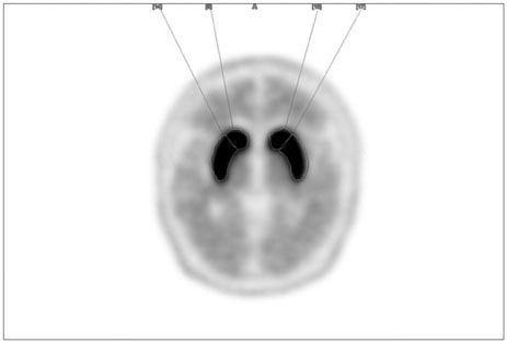
Fig. 1 Applications of the region of interest to 18F-FP-CIT positron emission tomography imaging showing right and left caudate, putamen at the level of striatum in a healthy control.
Reference
-
1. Fearnley JM, Lees AJ. Ageing and Parkinson's disease: substantia nigra regional selectivity. Brain. 1991; 114:2283–2301.
Article2. Kaufman MJ, Madras BK. Severe depletion of cocaine recognition sites associated with the dopamine transporter in Parkinson's-diseased striatum. Synapse. 1991; 9:43–49.
Article3. Huang WS, Chiang YH, Lin JC, Chou YH, Cheng CY, Liu RS. Crossover study of (99m)Tc-TRODAT-1 SPECT and (18)F-FDOPA PET in Parkinson's disease patients. J Nucl Med. 2003; 44:999–1005.4. Rinne OJ, Nurmi E, Ruottinen HM, Bergman J, Eskola O, Solin O. [(18)F]FDOPA and [(18)F]CFT are both sensitive PET markers to detect presynaptic dopaminergic hypofunction in early Parkinson's disease. Synapse. 2001; 40:193–200.
Article5. Kazumata K, Dhawan V, Chaly T, Antonini A, Margouleff C, Belakhlef A, et al. Dopamine transporter imaging with fluorine-18-FP-CIT and PET. J Nucl Med. 1998; 39:1521–1530.6. Robeson W, Dhawan V, Belakhlef A, Ma Y, Pillai V, Chaly T, et al. Dosimetry of the dopamine transporter radioligand 18F-FPCIT in human subjects. J Nucl Med. 2003; 44:961–966.7. Ma Y, Dhawan V, Mentis M, Chaly T, Spetsieris PG, Eidelberg D. Parametric mapping of [18F]FPCIT binding in early stage Parkinson's disease: a PET study. Synapse. 2002; 45:125–133.
Article8. Rinne JO, Ruottinen H, Bergman J, Haaparanta M, Sonninen P, Solin O. Usefulness of a dopamine transporter PET ligand [(18)F]beta-CFT in assessing disability in Parkinson's disease. J Neurol Neurosurg Psychiatry. 1999; 67:737–741.
Article9. Nurmi E, Ruottinen HM, Kaasinen V, Bergman J, Haaparanta M, Solin O, et al. Progression in Parkinson's disease: a positron emission tomography study with a dopamine transporter ligand [18F]CFT. Ann Neurol. 2000; 47:804–808.
Article10. Nurmi E, Bergman J, Eskola O, Solin O, Vahlberg T, Sonninen P, et al. Progression of dopaminergic hypofunction in striatal subregions in Parkinson's disease using [18F]CFT PET. Synapse. 2003; 48:109–115.
Article11. Chung M, Park YS, Kim JS, Kim YJ, Ma HI, Jang SJ, et al. Correlating Parkinson's disease motor symptoms with three-dimensional [(18)F]FP-CIT PET. Jpn J Radiol. 2015; 33:609–618.
Article12. Benamer HT, Patterson J, Wyper DJ, Hadley DM, Macphee GJ, Grosset DG. Correlation of Parkinson's disease severity and duration with 123I-FP-CIT SPECT striatal uptake. Mov Disord. 2000; 15:692–698.
Article13. Hughes AJ, Daniel SE, Kilford L, Lees AJ. Accuracy of clinical diagnosis of idiopathic Parkinson's disease: a clinico-pathological study of 100 cases. J Neurol Neurosurg Psychiatry. 1992; 55:181–184.
Article14. Lewis SJ, Foltynie T, Blackwell AD, Robbins TW, Owen AM, Barker RA. Heterogeneity of Parkinson's disease in the early clinical stages using a data driven approach. J Neurol Neurosurg Psychiatry. 2005; 76:343–348.
Article15. Kang YW, Na DL, Hahn SH. A validity study on the Korean minimental state examination (K-MMSE) in dementia patients. J Korean Neurol Assoc. 1997; 15:300–308.16. Isaias IU, Benti R, Cilia R, Canesi M, Marotta G, Gerundini P, et al. [123I]FP-CIT striatal binding in early Parkinson's disease patients with tremor vs. akinetic-rigid onset. Neuroreport. 2007; 18:1499–1502.
Article17. Nandhagopal R, Kuramoto L, Schulzer M, Mak E, Cragg J, Lee CS, et al. Longitudinal progression of sporadic Parkinson's disease: a multitracer positron emission tomography study. Brain. 2009; 132:2970–2979.
Article18. Wang J, Zuo CT, Jiang YP, Guan YH, Chen ZP, Xiang JD, et al. 18F-FP-CIT PET imaging and SPM analysis of dopamine transporters in Parkinson's disease in various Hoehn & Yahr stages. J Neurol. 2007; 254:185–190.
Article19. Marek K, Innis R, van Dyck C, Fussell B, Early M, Eberly S, et al. [123I]beta-CIT SPECT imaging assessment of the rate of Parkinson's disease progression. Neurology. 2001; 57:2089–2094.
Article20. Lee CS, Samii A, Sossi V, Ruth TJ, Schulzer M, Holden JE, et al. In vivo positron emission tomographic evidence for compensatory changes in presynaptic dopaminergic nerve terminals in Parkinson's disease. Ann Neurol. 2000; 47:493–503.
Article21. Hilker R, Schweitzer K, Coburger S, Ghaemi M, Weisenbach S, Jacobs AH, et al. Nonlinear progression of Parkinson disease as determined by serial positron emission tomographic imaging of striatal fluorodopa F 18 activity. Arch Neurol. 2005; 62:378–382.
Article22. Vingerhoets FJ, Schulzer M, Calne DB, Snow BJ. Which clinical sign of Parkinson's disease best reflects the nigrostriatal lesion? Ann Neurol. 1997; 41:58–64.
Article23. Hubbuch M, Farmakis G, Schaefer A, Behnke S, Schneider S, Hellwig D, et al. FP-CIT SPECT does not predict the progression of motor symptoms in Parkinson's disease. Eur Neurol. 2011; 65:187–192.
Article24. Spiegel J, Hellwig D, Samnick S, Jost W, Möllers MO, Fassbender K, et al. Striatal FP-CIT uptake differs in the subtypes of early Parkinson's disease. J Neural Transm. 2007; 114:331–335.
Article25. Djaldetti R, Treves TA, Ziv I, Melamed E, Lampl Y, Lorberboym M. Use of a single [123I]-FP-CIT SPECT to predict the severity of clinical symptoms of Parkinson disease. Neurol Sci. 2009; 30:301–305.
Article26. Kahraman D, Eggers C, Schicha H, Timmermann L, Schmidt M. Visual assessment of dopaminergic degeneration pattern in 123I-FP-CIT SPECT differentiates patients with atypical parkinsonian syndromes and idiopathic Parkinson's disease. J Neurol. 2012; 259:251–260.
Article27. Deuschl G, Raethjen J, Baron R, Lindemann M, Wilms H, Krack P. The pathophysiology of parkinsonian tremor: a review. J Neurol. 2000; 247:Suppl 5. V33–V48.
Article28. Lalley PM, Rossi GV, Baker WW. Tremor induction by intracaudate injections of bretylium, tetrabenazine, or mescaline: functional deficits in caudate dopamine. J Pharm Sci. 1973; 62:1302–1307.
Article29. Gelb DJ, Oliver E, Gilman S. Diagnostic criteria for Parkinson disease. Arch Neurol. 1999; 56:33–39.
Article30. Brück A, Aalto S, Rauhala E, Bergman J, Marttila R, Rinne JO. A follow-up study on 6-[18F]fluoro-L-dopa uptake in early Parkinson's disease shows nonlinear progression in the putamen. Mov Disord. 2009; 24:1009–1015.
Article31. Song IU, Kim YD, Cho HJ, Chung SW, Chung YA. An FP-CIT PET comparison of the differences in dopaminergic neuronal loss between idiopathic Parkinson disease with dementia and without dementia. Alzheimer Dis Assoc Disord. 2013; 27:51–55.
Article
- Full Text Links
-
- Actions
-
Cited
- CITED
-
- Close
- Share
-
- Similar articles
-
- Practical Approach for the Clinical Use of Dopamine Transporter Imaging
- Synthesis of a Dopamine Transporter Imaging Agent, N-(3-[18F]fluoropropyl)-2 -carbomethoxy-3 -(4-iodophenyl)nortropane
- Imaging Procedure and Clinical Studies of [18 F]FP‑CIT PET
- Salivary Gland Uptake on 18F-FP-CIT PET as a New Biomarker in Patients With Parkinsonism
- The Effect of SSRIs on the Binding of 18F-FP-CIT in Parkinson Patients: A Retrospective Case Control Study
