Duplicated Internal Auditory Canal: High-Resolution CT and MRI Findings
- Affiliations
-
- 1Imaging Center, the Affiliated Hospital of Jining Medical University, Jining, China.
- 2Department of Radiology, the First People's Hospital of Jining City, Jining, China. zlh968968@163.com
- KMID: 2442716
- DOI: http://doi.org/10.3348/kjr.2018.0065
Abstract
OBJECTIVE
To summarize the high-resolution computed tomography (HRCT) and magnetic resonance imaging (HRMRI) features of duplicated internal auditory canals (DIACs).
MATERIALS AND METHODS
Ear HRCT data of 64813 patients with sensorineural hearing loss (SNHL), obtained between August 2009 and November 2017, were reviewed. Among these patients, 12 (13 ears) were found to have DIACs, 9 of whom underwent HRMRI. Their images were evaluated by two otoradiologists.
RESULTS
The rate of occurrence of DIAC among SNHL patients was 0.019% (12/64813). The internal auditory canals of 13 ears were divided into double canals by complete (n = 6) and incomplete (n = 7) bony septa, with varied orientations ranging from horizontal to approximately vertical. All of the anterosuperior canals extended into the facial nerve (FN) canal, except for 1, which also extended to the vestibule. The posteroinferior canals ended in the cochlea and vestibule, except for 2, which also connected to the FN canals. Magnetic resonance images revealed that 77.8% (7/9) and 22.2% (2/9) of vestibulocochlear nerves (VCNs) were aplastic and hypoplastic, respectively. Furthermore, 88.9% (8/9) of FNs were normal, except for 1, which was hypoplastic. All of the affected ears also had other ear anomalies: a narrow, bony cochlear nerve canal was the most common other anomaly, accounting for 92.3% (12/13). Malformations of other systems were not found.
CONCLUSION
Double-canal appearance is a characteristic finding of DIAC on HRCT, and it is usually accompanied by other ear anomalies. The VCN usually appears aplastic, with a normal FN, on HRMRI.
MeSH Terms
Figure
-
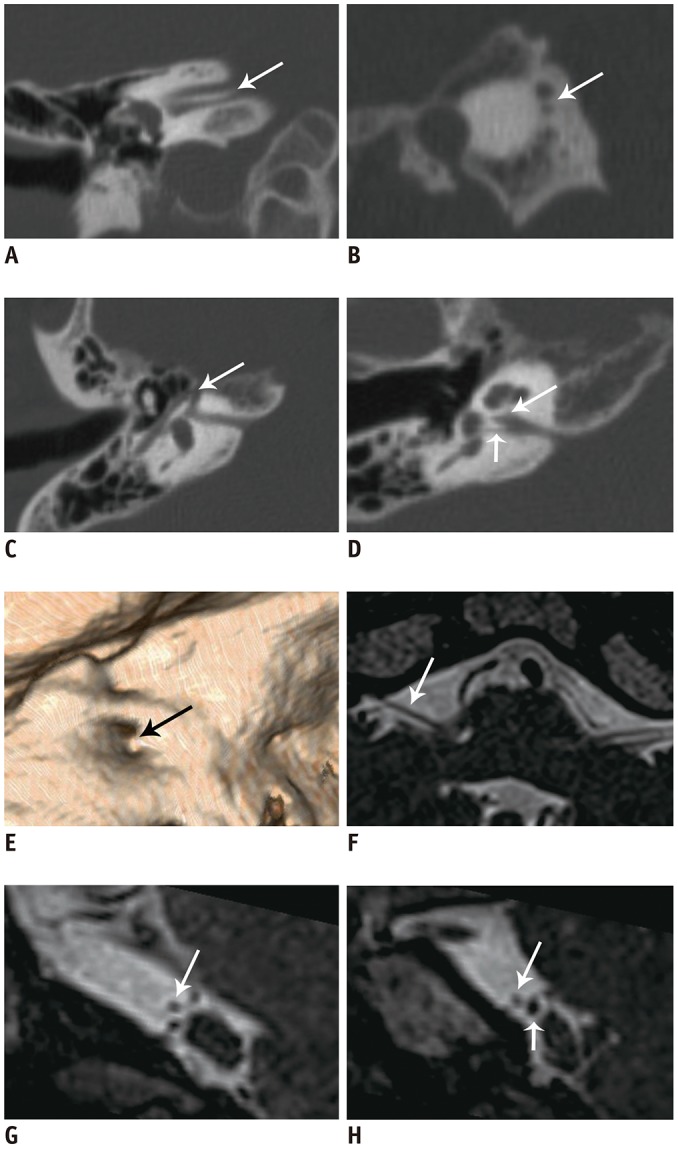
Fig. 1 23-year-old male with right DIAC.A, B. Oblique coronal and parasagittal HRCT images show that IAC is divided into double canals by complete horizontal bony septum (arrows). C, D. Superior portion is connected to FN canal (arrows), whereas inferior portion is connected to cochlea and vestibule (short arrow). Ipsilateral bony CN canal is narrow (long arrow). E. CT volume rendering image shows bony septum (arrow). F–H. Oblique axial and parasagittal MRI show that right VCN (short arrow) is aplastic and FN (long arrows) is normal in cisternal segment. CN = cochlear nerve, CT = computed tomography, DIAC = duplicated internal auditory canal, FN = facial nerve, HRCT = high-resolution CT, IAC = internal auditory canal, VCN = vestibulocochlear nerve
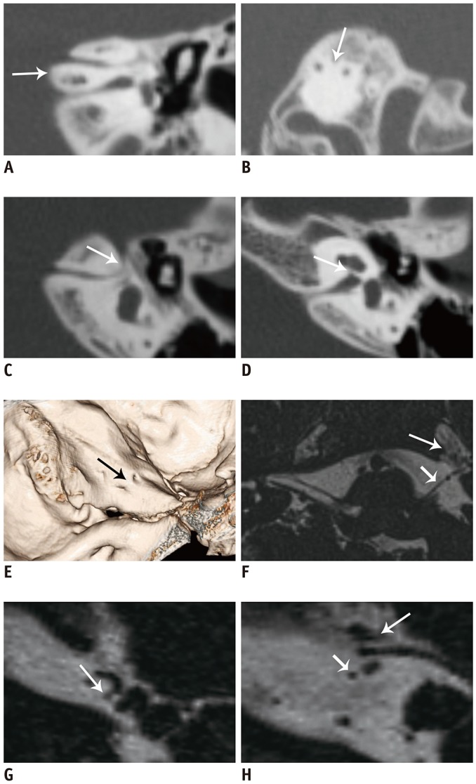
Fig. 2 6-year-old female with left DIAC.A, B. Oblique coronal and parasagittal HRCT images clearly show complete and nearly vertical bony septum (arrows). C. Anterior canal is connected to FN canal (arrow). D. Posterior canal ends in cochlea and vestibule, and ipsilateral bony CN canal is stenotic (arrow). E. CT volume rendering image clearly shows that FN canal meatus is anteriorly and superiorly located (arrow). F–H. Oblique axial and parasagittal MR images reveal left aplastic VCN and normal FN (short arrows). Latter has migrated beneath trigeminal nerve (long arrows). MR = magnetic resonance
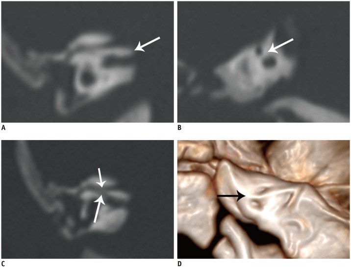
Fig. 3 4-month-old female with right DIAC.A, B. Oblique coronal and parasagittal HRCT images show that IAC is divided into two portions by complete oblique bony septum (arrows). C. FN canal is continuous with anterosuperior canal (short arrow) and with posteroinferior canal through accessory canal (long arrow). D. CT volume rendering image shows meatus of double canals and bony septum (arrow).
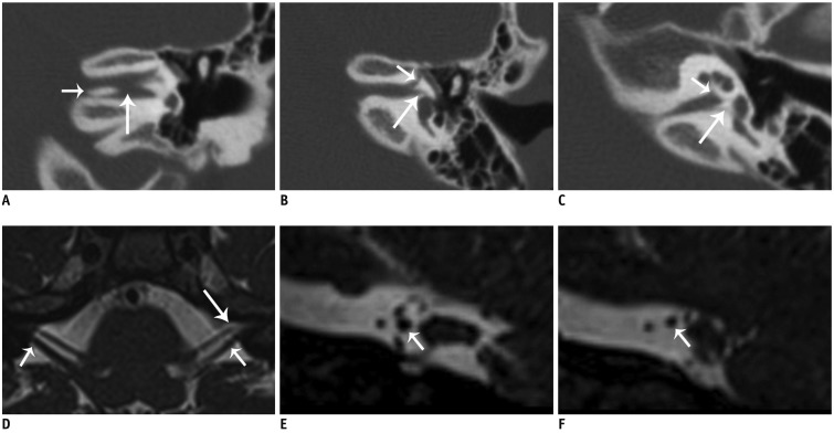
Fig. 4 8-year-old female with left DIAC.A. Oblique coronal HRCT image shows that bony septum has medial (short arrow) and lateral (long arrow) defects. B. Anterosuperior canal is connected to FN canal (short arrow) and to vestibule (long arrow). C. Posteroinferior canal ends in cochlea (short arrow) and vestibule (long arrow). D–F. Oblique axial and parasagittal MR images reveal that left dysplastic VCN (short arrows) and normal FN enter anterosuperior canal (long arrow).
Reference
-
1. Clemens F, Sandström J. Double-barreled hypoplastic internal auditory canal in unilateral deafness. Acta Radiol Diagn (Stockh). 1975; 16:342–346. PMID: 1189959.
Article2. Goktas Bakar T, Karadag D, Calisir C, Adapinar B. Bilateral narrow duplicated internal auditory canal. Eur Arch Otorhinolaryngol. 2008; 265:999–1001. PMID: 18176812.
Article3. Curtin H, May M. Double internal auditory canal associated with progressive facial weakness. Am J Otol. 1986; 7:275–281. PMID: 3740236.4. Weissman JL, Arriaga M, Curtin HD, Hirsch B. Duplication anomaly of the internal auditory canal. AJNR Am J Neuroradiol. 1991; 12:867–869. PMID: 1950913.5. Weon YC, Kim JH, Choi SK, Koo JW. Bilateral duplication of the internal auditory canal. Pediatr Radiol. 2007; 37:1047–1049. PMID: 17704912.
Article6. Ferreira T, Shayestehfar B, Lufkin R. Narrow, duplicated internal auditory canal. Neuroradiology. 2003; 45:308–310. PMID: 12743665.
Article7. Cho YS, Na DG, Jung JY, Hong SH. Narrow internal auditory canal syndrome: parasagittal reconstruction. J Laryngol Otol. 2000; 114:392–394. PMID: 10912275.8. Baik HW, Yu H, Kim KS, Kim GH. A narrow internal auditory canal with duplication in a patient with congenital sensorineural hearing loss. Korean J Radiol. 2008; 9(Suppl):S22–S25. PMID: 18607120.
Article9. Casselman JW, Offeciers FE, Govaerts PJ, Kuhweide R, Geldof H, Somers T, et al. Aplasia and hypoplasia of the vestibulocochlear nerve: diagnosis with MR imaging. Radiology. 1997; 202:773–781. PMID: 9051033.
Article10. Vilain J, Pigeolet Y, Casselman JW. Narrow and vacant internal auditory canal. Acta Otorhinolaryngol Belg. 1999; 53:67–71. PMID: 10102042.11. Demir OI, Cakmakci H, Erdag TK, Men S. Narrow duplicated internal auditory canal: radiological findings and review of the literature. Pediatr Radiol. 2005; 35:1220–1223. PMID: 16079980.
Article12. Kesser BW, Raghavan P, Mukherjee S, Carfrae M, Essig G, Hashisaki GT. Duplication of the internal auditory canal: radiographic imaging case of the month. Otol Neurotol. 2010; 31:1352–1353. PMID: 20864882.13. Ramírez-Camacho R, Berrocal JR, Arellano B. Bilateral malformation of the internal auditory canal: atresia and contralateral transverse megacrest. Otolaryngol Head Neck Surg. 2001; 125:115–116. PMID: 11458231.
Article14. Guirado CR. Malformations of the inner auditory canal. Rev Laryngol Otol Rhinol (Bord). 1992; 13:419–421.15. Lee SY, Cha SH, Jeon MH, Bae IH, Han GS, Kim SJ, et al. Narrow duplicated or triplicated internal auditory canal (3 cases and review of literature): can we regard the separated narrow internal auditory canal as the presence of vestibulocochlear nerve fibers? J Comput Assist Tomogr. 2009; 33:565–570. PMID: 19638851.16. Vincenti V, Ormitti F, Ventura E. Partitioned versus duplicated internal auditory canal: when appropriate terminology matters. Otol Neurotol. 2014; 35:1140–1144. PMID: 24836591.17. Kono T, Kuwashima S, Arakawa H, Yamazaki E, Kitajima K, Ejima Y, et al. Narrow duplicated internal auditory canal: a rare inner ear malformation with sensorineural hearing loss. Arch Otolaryngol Head Neck Surg. 2009; 135:1048–1051. PMID: 19841348.18. Kew TY, Abdullah A. Duplicate internal auditory canals with facial and vestibulocochlear nerve dysfunction. J Laryngol Otol. 2012; 126:66–71. PMID: 21867589.
Article19. Kishimoto I, Moroto S, Fujiwara K, Harada H, Kikuchi M, Suehiro A, et al. Bilateral duplication of the internal auditory canal: a case with successful cochlear implantation. Int J Pediatr Otorhinolaryngol. 2015; 79:1595–1598. PMID: 26209350.
Article20. Binnetoğlu A, Bağlam T, Sarı M, Gündoğdu Y, Batman Ç. A challenge for cochlear implantation: duplicated internal auditory canal. J Int Adv Otol. 2016; 12:199–201. PMID: 27716607.21. Sakina MS, Goh BS, Abdullah A, Zulfiqar MA, Saim L. Internal auditory canal stenosis in congenital sensorineural hearing loss. Int J Pediatr Otorhinolaryngol. 2006; 70:2093–2097. PMID: 16996619.
Article22. Desai NK, Young L, Miranda MA, Kutz JW Jr, Roland PS, Booth TN. Pontine tegmental cap dysplasia: the neurotologic perspective. Otolaryngol Head Neck Surg. 2011; 145:992–998. PMID: 21705787.23. Nixon JN, Dempsey JC, Doherty D, Ishak GE. Temporal bone and cranial nerve findings in pontine tegmental cap dysplasia. Neuroradiology. 2016; 58:179–187. PMID: 26458891.
Article24. Wang LS, Zhang LH, Sun XH, Yang YY, Chen YQ, Li X, et al. [Imaging features of duplication of the internal auditory canal.]. Zhonghua Er Bi Yan Hou Tou Jing Wai Ke Za Zhi. 2010; 45:481–485. PMID: 21055326.25. Wang LS, Zhang LH, Li XY, Chen YQ, Li X, Sun ZG, et al. [CT and MRI diagnosis of congenital stenosis of the internal auditory canal.]. Zhonghua Er Bi Yan Hou Tou Jing Wai Ke Za Zhi. 2011; 46:533–538. PMID: 22088279.26. Casselman JW, Offeciers EF, De Foer B, Govaerts P, Kuhweide R, Somers T. CT and MR imaging of congenital abnormalities of the inner ear and internal auditory canal. Eur J Radiol. 2001; 40:94–104. PMID: 11704356.27. Norton SJ, Gorga MP, Widen JE, Folsom RC, Sininger Y, Cone-Wesson B, et al. Identification of neonatal hearing impairment: summary and recommendations. Ear Hear. 2000; 21:529–535. PMID: 11059708.
Article28. Sennaroglu L, Saatci I. A new classification for cochleovestibular malformations. Laryngoscope. 2002; 112:2230–2241. PMID: 12461346.
Article29. Stjernholm C, Muren C. Dimensions of the cochlear nerve canal: a radioanatomic investigation. Acta Otolaryngol. 2002; 122:43–48. PMID: 11876597.
Article
- Full Text Links
-
- Actions
-
Cited
- CITED
-
- Close
- Share
-
- Similar articles
-
- The Significance of a Hypoplastic Bony Canal for the Cochlear Nerve in Patients with Sensorineural Hearing Loss: CT and MRI Findings
- Nerve Canals at the Fundus of the Internal Auditory Canal on High-Resolution Temporal Bone CT
- A case report of narrow internal auditory canal: Parasagittal reconstruction
- A Case of Solitary Fibrous Tumor in the External Auditory Canal
- CT Findings of the Osteoma of the External Auditory Canal
