Korean J Ophthalmol.
2019 Apr;33(2):167-172. 10.3341/kjo.2018.0099.
Measurement of Contrast Sensitivity in Patients with Behçet's Disease without Ocular Involvement
- Affiliations
-
- 1Department of Ophthalmology, Haseki Training and Research Hospital, University of Health Sciences, Istanbul, Turkey. ercanozy@hotmail.com
- 2Department of Ophthalmology, Inonu University School of Medicine, Malatya, Turkey.
- KMID: 2442621
- DOI: http://doi.org/10.3341/kjo.2018.0099
Abstract
- PURPOSE
To evaluate contrast sensitivity in patients with Behçet's disease (BD) without ocular involvement.
METHODS
The study group was composed of 47 BD patients (20 to 50 years of age) who did not have ocular involvement. The control group was composed of 47 normal volunteers who were similar to the study group in terms of age and gender. No participants in this study had any ocular or systemic pathologies except for BD. The contrast sensitivity measurements were performed using the Functional Acuity Contrast Test under photopic conditions, and the results were compared between the two groups.
RESULTS
The mean age of the BD patients and control subjects was 34.5 ± 9.7 and 33.2 ± 7.6 years, respectively. The mean disease duration of the BD patients was 5.5 ± 6.4 years. There was a statistically significant decrease at five spatial frequencies (A, 1.5; B, 3; C, 6; D, 2; and E, 18 cycles per degree) in patients with BD compared with control subjects (p < 0.001, p = 0.004, p = 0.002, p < 0.001, and p = 0.001, respectively).
CONCLUSIONS
The contrast sensitivity of BD patients without ocular involvement was lower than that of the control group. Further studies seem mandatory to confirm our results.
Keyword
Figure
-
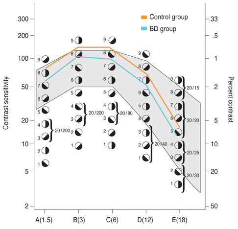
Fig. 1 The contrast sensitivity of the groups at five spatial frequencies. BD = Behçet's disease; cpd = cycles per degree.
Reference
-
1. Yazici H, Yurdakul S, Hamuryudan V. Behcet's syndrome. Curr Opin Rheumatol. 1999; 11:53–57.2. Al-Dhibi H, Abouammoh M, Al-Harthi E, et al. Macular hole in Behcet's disease. Indian J Ophthalmol. 2011; 59:359–362.3. Criteria for diagnosis of Behcet's disease. International Study Group for Behcet's Disease. Lancet. 1990; 335:1078–1080.4. A SE, Moses PD, George R. Ocular manifestations of Behcet's disease. Indian Pediatr. 2005; 42:942–945.5. Borhani Haghighi A, Sharifzad HR, Matin S, Rezaee S. The pathological presentations of neuro-Behcet disease: a case report and review of the literature. Neurologist. 2007; 13:209–214.6. Saylan T, Mat C, Fresko I, Melikoglu M. Behcet's disease in the Middle East. Clin Dermatol. 1999; 17:209–223.7. Yazici H, Fresko I, Yurdakul S. Behcet's syndrome: disease manifestations, management, and advances in treatment. Nat Clin Pract Rheumatol. 2007; 3:148–155.8. Yuksel H, Turkcu FM, Sahin M, et al. Inner and outer segment junction (IS/OS line) integrity in ocular Behcet's disease. Arq Bras Oftalmol. 2014; 77:219–221.9. Arden GB. The importance of measuring contrast sensitivity in cases of visual disturbance. Br J Ophthalmol. 1978; 62:198–209.
Article10. Olsen T, Corydon L. Contrast sensitivity as a function of focus in patients with the diffractive multifocal intraocular lens. J Cataract Refract Surg. 1990; 16:703–706.
Article11. Ginsburg AP. Contrast sensitivity: determining the visual quality and function of cataract, intraocular lenses and refractive surgery. Curr Opin Ophthalmol. 2006; 17:19–26.12. Loshin DS, White J. Contrast sensitivity: the visual rehabilitation of the patient with macular degeneration. Arch Ophthalmol. 1984; 102:1303–1306.13. Trick GL, Burde RM, Gordon MO, et al. The relationship between hue discrimination and contrast sensitivity deficits in patients with diabetes mellitus. Ophthalmology. 1988; 95:693–698.
Article14. Marmor MF. Contrast sensitivity versus visual acuity in retinal disease. Br J Ophthalmol. 1986; 70:553–559.
Article15. Ridder A, Muller ML, Kotagal V, et al. Impaired contrast sensitivity is associated with more severe cognitive impairment in Parkinson disease. Parkinsonism Relat Disord. 2017; 34:15–19.
Article16. Preti RC, Ramirez LM, Monteiro ML, et al. Contrast sensitivity evaluation in high risk proliferative diabetic retinopathy treated with panretinal photocoagulation associated or not with intravitreal bevacizumab injections: a randomised clinical trial. Br J Ophthalmol. 2013; 97:885–889.
Article17. Klaeger A, Tran VT, Hiroz CA, et al. Indocyanine green angiography in Behcet's uveitis. Retina. 2000; 20:309–314.
Article18. Gedik S, Akova Y, Yilmaz G, Bozbeyoglu S. Indocyanine green and fundus fluorescein angiographic findings in patients with active ocular Behcet's disease. Ocul Immunol Inflamm. 2005; 13:51–58.19. Coskun E, Gurler B, Pehlivan Y, et al. Enhanced depth imaging optical coherence tomography findings in Behcet disease. Ocul Immunol Inflamm. 2013; 21:440–445.20. Farahangiz S, Sarhadi S, Safari A, Borhani-Haghighi A. Magnetic resonance imaging findings and outcome of neuro-Behcet's disease: the predictive factors. Int J Rheum Dis. 2012; 15:e142–e149.21. Borman P, Tuncay F, Kocaoglu S, et al. The subclinic autonomic dysfunction in patients with Behcet disease: an electrophysiological study. Clin Rheumatol. 2012; 31:41–47.22. Hirohata S, Kikuchi H, Sawada T, et al. Retrospective analysis of long-term outcome of chronic progressive neurological manifestations in Behcet's disease. J Neurol Sci. 2015; 349:143–148.
Article23. van Ham C, Schrijvers D, De Picker L, et al. Neuropsychiatric features in Behcet's disease: a case report. Clin Neurol Neurosurg. 2014; 127:13–14.24. Safi S, Rahimi A, Raeesi A, et al. Contrast sensitivity to spatial gratings in moderate and dim light conditions in patients with diabetes in the absence of diabetic retinopathy. BMJ Open Diabetes Res Care. 2017; 5:e000408.
Article25. Safi H, Safi S, Hafezi-Moghadam A, Ahmadieh H. Early detection of diabetic retinopathy. Surv Ophthalmol. 2018; 63:601–608.
Article26. Di Leo MA, Caputo S, Falsini B, et al. Nonselective loss of contrast sensitivity in visual system testing in early type I diabetes. Diabetes Care. 1992; 15:620–625.
Article27. Holland GN, Kappel PJ, Van Natta ML, et al. Association between abnormal contrast sensitivity and mortality among people with acquired immunodeficiency syndrome. Am J Ophthalmol. 2010; 149:807–816.
Article28. Owsley C, Sekuler R, Siemsen D. Contrast sensitivity throughout adulthood. Vision Res. 1983; 23:689–699.
Article29. Volkers AC, Hagemans KH, van der Wildt GJ, Schmitz PI. Spatial contrast sensitivity and the diagnosis of amblyopia. Br J Ophthalmol. 1987; 71:58–65.
Article30. Nomura H, Ando F, Niino N, et al. Age-related change in contrast sensitivity among Japanese adults. Jpn J Ophthalmol. 2003; 47:299–303.
Article
- Full Text Links
-
- Actions
-
Cited
- CITED
-
- Close
- Share
-
- Similar articles
-
- Rupture of Renal Artery in a Patient with Behçet's Disease
- Behçet's Disease with Deep Vaginal Ulcer Diagnosed for the First Time during Pregnancy
- A Study of Interleukin-2 Activity in Behçet's Syndrome
- Blepharoptosis in Behçet's Disease
- Successfully treated isolated renal artery pseudoaneurysm in a patient with Behçet's disease
