Ectopic Mediastinal Parathyroid Adenoma in a Patient with Chronic Kidney Disease: A Case Report
- Affiliations
-
- 1Department of Radiology, Chungnam National University Hospital, Chungnam National University School of Medicine, Daejeon, Korea. michelan@cnu.ac.kr
- KMID: 2442475
- DOI: http://doi.org/10.3348/jksr.2019.80.1.147
Abstract
- We report an ectopic mediastinal parathyroid adenoma in a patient with chronic kidney disease who presented with refractory hypercalcemia. Technetium-99m-sestamibi scintigraphy is a valuable imaging technique for the detection of an ectopic parathyroid adenoma in the mediastinum. The combination of intense contrast enhancement and the identification of a polar vessel on a computed tomography scan will ensure that a radiologist can differentiate a parathyroid adenoma from other pathologies in the mediastinum. By virtue of the advancement of imaging modalities, localization of an ectopic mediastinal parathyroid adenoma prior to surgery is possible and unnecessary neck exploration is avoidable.
MeSH Terms
Figure
-
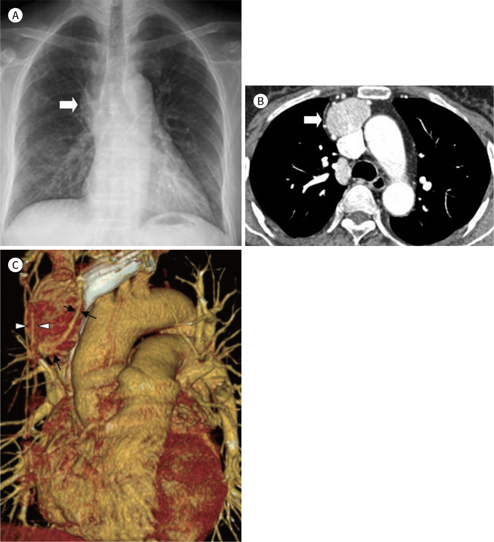
Fig. 1. An ectopic anterior mediastinal parathyroid adenoma in a 63-year-old woman. A. Initial posteroanterior chest radiograph shows widening with contour bulging of the right mediastinum (arrow). B. A contrast-enhanced chest computed tomography axial image shows a highly enhancing soft tissue mass in the right anterior mediastinum, measuring 4 × 3 cm. The mass is located anterior to the superior vena cava (arrow). C. Three-dimensional volume rendering reconstruction image demonstrates an enlarged tortuous feed-ing artery originating from the right internal mammary artery (arrowheads) and a dilated vein draining to the right internal mammary vein at the inferior pole of the tumor (arrows).

Fig. 1. An ectopic anterior mediastinal parathyroid adenoma in a 63-year-old woman. D. Early phase (left) and delayed phase (middle) coronaldual-phase Technetium-99m-sestamibi scintigraphy and fused single photon emission computed tomography/computed tomography (right) images depict a focal and persistent abnormal tracer accumulation in the right anterior mediastinal mass. E. Gross specimen photograph shows a resected ectopic parathyroid adenoma (4 cm). The tumor is well encapsulated and firmly adhered to fatty thy-mic tissue.
Reference
-
References
1. Eslamy HK, Ziessman HA. Parathyroid scintigraphy in patients with primary hyperparathyroidism: 99mTc sestamibi SPECT and SPECT/CT.Rad/iiograph/iics. 2008; 28:1461–1476.2. Cozzolino M. CKD-MBD KDIGO guidelines: how difficult is reaching the ‘target'? Clinical Kidney J. 2018; 11:70–72.
Article3. Yuen NK, Ananthakrishnan S, Campbell MJ. Hyperparathyroidism of renal disease.Perm J. 2016; 20:78–83.4. Lunn MR, Muñoz Mendoza J, Pasche LJ, Norton JA, Ayco AL, Chertow GM. Hyperparathyroidism with hypercalcaemia in chronic kidney disease: primary or tertiary? NDT Plus. 2010; 3:366–371.
Article5. Hu J, Ngiam KY, Parameswaran R. Mediastinal parathyroid adenomas and their surgical implications. Ann R Coll Surg Engl. 2015; 97:259–261.
Article6. Gordon J, Manley NR. Mechanisms of thymus organogenesis and morphogenesis. Development. 2011; 138:3865–3878.
Article7. Roy M, Mazeh H, Chen H, Sippel RS. Incidence and localization of ectopic parathyroid adenomas in previously unexplored patients. World J Surg. 2013; 37:102–106.
Article8. Lane MJ, Desser TS, Weigel RJ, Jeffrey RB Jr. Use of color and power Doppler sonography to identify feeding arteries associated with parathyroid adenomas.AJR Am J Roentgenol. 1998; 171:819–823.9. Rodgers SE, Hunter GJ, Hamberg LM, Schellingerhout D, Doherty DB, Ayers GD, et al. Improved preoperative planning for directed parathyroidectomy with 4-dimensional computed tomography.Surgery. 2006; 140:932–940. ; discussion 940–941.10. Bahl M, Muzaffar M, Vij G, Sosa JA, Choudhury KR, Hoang JK. Prevalence of the polar vessel sign in parathyroid adenomas on the arterial phase of 4D CT. AJNR Am J Neuroradiol. 2014; 35:578–581.
Article
- Full Text Links
-
- Actions
-
Cited
- CITED
-
- Close
- Share
-
- Similar articles
-
- Supernumerary Ectopic Mediastinal Parathyroid Adenoma Combined With Parathyroid Hyperplasia
- Thoracoscopic Removal of Ectopic Mediastinal Parathyroid Adenoma
- A Case of Mediastinal parathyroid adenoma localized by technetium-99m sestamibi scanning
- A Case of Mediastinal Cystic Parathyroid Adenoma Presenting as Acute Pancreatitis
- A Case of Hyperparathyroidism Caused by Intrathyroidal Parathyroid Adenoma
