J Breast Cancer.
2019 Mar;22(1):52-66. 10.4048/jbc.2019.22.e4.
Screening of Recurrence Related MicroRNA in Ductal Carcinoma In Situ and Functional Study of MicroRNA-654-5p
- Affiliations
-
- 1Department of Pathology, The Fourth Hospital of Hebei Medical University, Shijiazhuang, China. annama@163.com
- 2Department of Pathology, West China Hospital, Sichuan University, Chengdu, China.
- 3Laboratory of Pathology, West China Hospital, Sichuan University, Chengdu, China.
- 4Breast Cancer Center, Key Laboratory of Transplant Engineering and Immunology, Ministry of Health, West China Hospital, Chengdu, China.
- KMID: 2441851
- DOI: http://doi.org/10.4048/jbc.2019.22.e4
Abstract
- PURPOSE
Ductal carcinoma in situ (DCIS) contributes to 20%-30% of newly diagnosed cases of breast cancer in China. Although the breast cancer-specific mortality of DCIS is extremely low, a small proportion of DCIS patients still show relapse or metastasis, leading to poor prognosis. Little is known about the molecular mechanism for DCIS metastasis, partly due to the limited number of poor prognosis patients. This study analyzed the clinicopathological features and screened key microRNAs (miRNAs) contributing to local or distant recurrence.
METHODS
The clinicopathological features of DCIS were evaluated and survival analysis were performed to clarify risk factors associated with poor prognosis. Using miRNA arrays and real-time quantitative polymerase chain reaction (RT-qPCR) on DCIS formalin-fixed and paraffin-embedded samples with or without microinvasion with different clinical outcomes, potential DCIS metastasis-related miRNAs were screened out and further validated. The influence of one identified miRNA, miRNA-654-5p, on DCIS progression was analyzed.
RESULTS
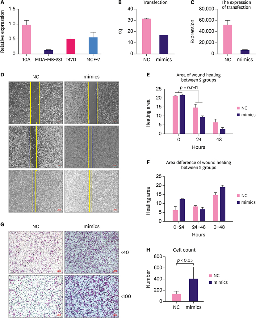
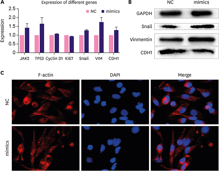
Poor prognosis was significantly associated with larger tumor size and higher lymph node metastasis rate (both p < 0.05). Both were independent prognostic factors for DCIS. According to RT-qPCR results, distinct miRNA expression profiles were identified between DCIS and DCIS with microinvasion (DCIS-Mi) patients. In the DCIS panel, miRNA-654-5p was significantly upregulated in the patients with poor prognosis. In vitro, miRNA-654-5p promoted MDA-MB-231 cell mobility in healing tests and metastasis in the Transwell study.
CONCLUSION
The panel of high-risk miRNAs in DCIS and DCIS-Mi differs markedly. miRNA-654-5p is significantly upregulated DCIS patients having poor prognosis and may be essential for local and distant recurrence in DCIS.
Keyword
MeSH Terms
Figure
Reference
-
1. Chen W, Zheng R, Baade PD, Zhang S, Zeng H, Bray F, et al. Cancer statistics in China, 2015. CA Cancer J Clin. 2016; 66:115–132.
Article2. Fan L, Zheng Y, Yu KD, Liu GY, Wu J, Lu JS, et al. Breast cancer in a transitional society over 18 years: trends and present status in Shanghai, China. Breast Cancer Res Treat. 2009; 117:409–416.
Article3. Narod SA, Iqbal J, Giannakeas V, Sopik V, Sun P. Breast cancer mortality after a diagnosis of ductal carcinoma in situ. JAMA Oncol. 2015; 1:888–896.
Article4. Rakovitch E, Mihai A, Pignol JP, Hanna W, Kwinter J, Chartier C, et al. Is expert breast pathology assessment necessary for the management of ductal carcinoma in situ? Breast Cancer Res Treat. 2004; 87:265–272.
Article5. Mardekian SK, Bombonati A, Palazzo JP. Ductal carcinoma in situ of the breast: the importance of morphologic and molecular interactions. Hum Pathol. 2016; 49:114–123.
Article6. Lee RC, Feinbaum RL, Ambros V. The C. elegans heterochronic gene lin-4 encodes small RNAs with antisense complementarity to lin-14. Cell. 1993; 75:843–854.
Article7. O'Bryan S, Dong S, Mathis JM, Alahari SK. The roles of oncogenic miRNAs and their therapeutic importance in breast cancer. Eur J Cancer. 2017; 72:1–11.8. Lewis BP, Burge CB, Bartel DP. Conserved seed pairing, often flanked by adenosines, indicates that thousands of human genes are microRNA targets. Cell. 2005; 120:15–20.
Article9. Hannafon BN, Sebastiani P, de las Morenas A, Lu J, Rosenberg CL. Expression of microRNA and their gene targets are dysregulated in preinvasive breast cancer. Breast Cancer Res. 2011; 13:R24.
Article10. Farazi TA, Horlings HM, Ten Hoeve JJ, Mihailovic A, Halfwerk H, Morozov P, et al. MicroRNA sequence and expression analysis in breast tumors by deep sequencing. Cancer Res. 2011; 71:4443–4453.
Article11. Lu M, Wang C, Chen W, Mao C, Wang J. miR-654-5p targets GRAP to promote proliferation, metastasis, and chemoresistance of oral squamous cell carcinoma through Ras/MAPK signaling. DNA Cell Biol. 2018; 37:381–388.
Article12. Mardekian SK, Bombonati A, Palazzo JP. Ductal carcinoma in situ of the breast: the importance of morphologic and molecular interactions. Hum Pathol. 2016; 49:114–123.
Article13. Hannafon BN, Sebastiani P, de las Morenas A, Lu J, Rosenberg CL. Expression of microRNA and their gene targets are dysregulated in preinvasive breast cancer. Breast Cancer Res. 2011; 13:R24.
Article14. Chen L, Li Y, Fu Y, Peng J, Mo MH, Stamatakos M, et al. Role of deregulated microRNAs in breast cancer progression using FFPE tissue. PLoS One. 2013; 8:e54213.
Article15. Li S, Meng H, Zhou F, Zhai L, Zhang L, Gu F, et al. MicroRNA-132 is frequently down-regulated in ductal carcinoma in situ (DCIS) of breast and acts as a tumor suppressor by inhibiting cell proliferation. Pathol Res Pract. 2013; 209:179–183.
Article16. Ueno K, Hirata H, Shahryari V, Deng G, Tanaka Y, Tabatabai ZL, et al. microRNA-183 is an oncogene targeting Dkk-3 and SMAD4 in prostate cancer. Br J Cancer. 2013; 108:1659–1667.
Article17. Zha R, Guo W, Zhang Z, Qiu Z, Wang Q, Ding J, et al. Genome-wide screening identified that miR-134 acts as a metastasis suppressor by targeting integrin beta1 in hepatocellular carcinoma. PLoS One. 2014; 9:e87665.18. Gregory PA, Bert AG, Paterson EL, Barry SC, Tsykin A, Farshid G, et al. The miR-200 family and miR-205 regulate epithelial to mesenchymal transition by targeting ZEB1 and SIP1. Nat Cell Biol. 2008; 10:593–601.
Article19. Li P, Xu T, Zhou X, Liao L, Pang G, Luo W, et al. Downregulation of miRNA-141 in breast cancer cells is associated with cell migration and invasion: involvement of ANP32E targeting. Cancer Med. 2017; 6:662–672.
Article20. Roy SS, Gonugunta VK, Bandyopadhyay A, Rao MK, Goodall GJ, Sun LZ, et al. Significance of PELP1/HDAC2/miR-200 regulatory network in EMT and metastasis of breast cancer. Oncogene. 2014; 33:3707–3716.
Article21. Östling P, Leivonen SK, Aakula A, Kohonen P, Mäkelä R, Hagman Z, et al. Systematic analysis of microRNAs targeting the androgen receptor in prostate cancer cells. Cancer Res. 2011; 71:1956–1967.
Article22. Paydas S, Acikalin A, Ergin M, Celik H, Yavuz B, Tanriverdi K. Micro-RNA (miRNA) profile in Hodgkin lymphoma: association between clinical and pathological variables. Med Oncol. 2016; 33:34.
Article23. Tan YY, Xu XY, Wang JF, Zhang CW, Zhang SC. MiR-654-5p attenuates breast cancer progression by targeting EPSTI1. Am J Cancer Res. 2016; 6:522–532.24. Volinia S, Galasso M, Sana ME, Wise TF, Palatini J, Huebner K, et al. Breast cancer signatures for invasiveness and prognosis defined by deep sequencing of microRNA. Proc Natl Acad Sci U S A. 2012; 109:3024–3029.
Article25. Whipple RA, Balzer EM, Cho EH, Matrone MA, Yoon JR, Martin SS. Vimentin filaments support extension of tubulin-based microtentacles in detached breast tumor cells. Cancer Res. 2008; 68:5678–5688.
Article26. Szynglarewicz B, Kasprzak P, Donizy P, Biecek P, Halon A, Matkowski R. Epithelial-mesenchymal transition inducer Snail1 and invasive potential of intraductal breast cancer. J Surg Oncol. 2017; 116:696–705.
Article27. Scribner KC, Behbod F, Porter WW. Regulation of DCIS to invasive breast cancer progression by Singleminded-2s (SIM2s). Oncogene. 2013; 32:2631–2639.
Article28. Polioudaki H, Agelaki S, Chiotaki R, Politaki E, Mavroudis D, Matikas A, et al. Variable expression levels of keratin and vimentin reveal differential EMT status of circulating tumor cells and correlation with clinical characteristics and outcome of patients with metastatic breast cancer. BMC Cancer. 2015; 15:399.
Article29. Szynglarewicz B, Kasprzak P, Donizy P, Biecek P, Halon A, Matkowski R. Epithelial-mesenchymal transition inducer Snail1 and invasive potential of intraductal breast cancer. J Surg Oncol. 2017; 116:696–705.
Article
- Full Text Links
- Actions
-
Cited
- CITED
-
- Close
- Share
- Similar articles
-
- MicroRNA-98-5p Inhibits Tumorigenesis of Hepatitis B Virus-Related Hepatocellular Carcinoma by Targeting NF-κB-Inducing Kinase
- MicroRNA-139-5p Regulates Fibrotic Potentials via Modulation of Collagen Type 1 and Phosphorylated p38 MAPK in Uterine Leiomyoma
- An Unusual Presentation of Extensive Ductal Carcinoma in Situ Accompanying Invasive Ductal Carcinoma on MRI: A Case Report
- Invasive Lobular Carcinoma of the Breast Associated with Mixed Lobular and Ductal Carcinoma In Situ: A Case Report
- The Long Noncoding RNA DUXAP8 Facilitates the Malignant Progression of Colon Cancer via the microRNA-378a-3p/FOXQ1 Axis

