Korean J Orthod.
2018 Jan;48(1):3-10. 10.4041/kjod.2018.48.1.3.
Prediction of optimal bending angles of a running loop to achieve bodily protraction of a molar using the finite element method
- Affiliations
-
- 1Private Practice, Daejeon, Korea.
- 2Postgraduate Orthodontic Program, Arizona School of Dentistry & Oral Health, A. T. Still University, Mesa, AZ, USA. jongmoon@wku.ac.kr
- 3Graduate School of Dentistry, Kyung Hee University, Seoul, Korea.
- 4Private Practice, Okayama, Japan.
- 5Department of Mechanical Engineering, Nagoya Institute of Technology, Nagoya, Japan.
- 6Department of Orthodontics, Wonkwang University School of Dentistry, Iksan, Korea.
- 7Wonkwang Dental Research Institute, Wonkwang University School of Dentistry, Iksan, Korea.
- 8The Korean Orthodontic Research Institute Inc., Seoul, Korea.
- KMID: 2441436
- DOI: http://doi.org/10.4041/kjod.2018.48.1.3
Abstract
OBJECTIVE
The purpose of this study was to predict the optimal bending angles of a running loop for bodily protraction of the mandibular first molars and to clarify the mechanics of molar tipping and rotation.
METHODS
A three-dimensional finite element model was developed for predicting tooth movement, and a mechanical model based on the beam theory was constructed for clarifying force systems.
RESULTS
When a running loop without bends was used, the molar tipped mesially by 9.6° and rotated counterclockwise by 5.4°. These angles were almost similar to those predicted by the beam theory. When the amount of tip-back and toe-in angles were 11.5° and 9.9°, respectively, bodily movement of the molar was achieved. When the bend angles were increased to 14.2° and 18.7°, the molar tipped distally by 4.9° and rotated clockwise by 1.5°.
CONCLUSIONS
Bodily movement of a mandibular first molar was achieved during protraction by controlling the tip-back and toe-in angles with the use of a running loop. The beam theory was effective for understanding the mechanics of molar tipping and rotation, as well as for predicting the optimal bending angles.
Figure
-

Figure 1 Finite element model for simulating orthodontic tooth movement.
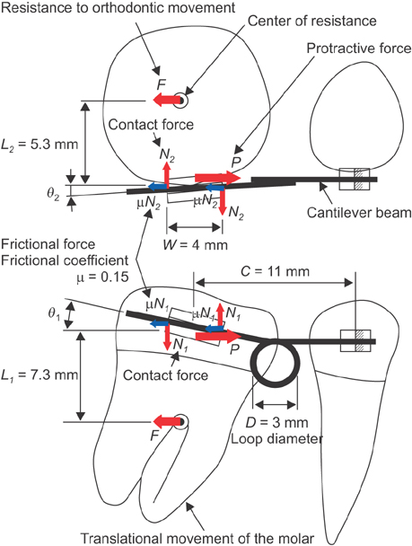
Figure 2 Forces acting on the molar under translational movement.
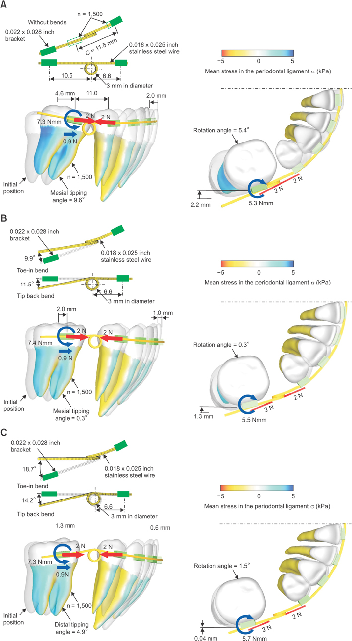
Figure 3 Effect of bending angles of the running loop on the tooth movement pattern. A, Without bends; B, tip-back angle (11.5°), toe-in angle (9.9°); C, tip-back angle (14.2°), toe-in angle (18.7°).
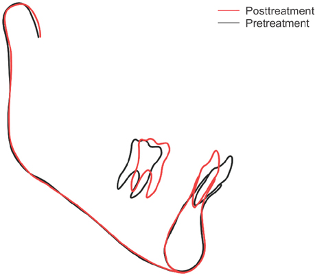
Figure 4 Tooth movement produced by the running loop used in clinical treatment.
Reference
-
1. Proffit WR. Forty-year review of extraction frequencies at a university orthodontic clinic. Angle Orthod. 1994; 64:407–414.2. Schoppe RJ. An Analysis of second premolar extraction procedures. Angle Orthod. 1964; 34:292–302.3. Brandt S, Safirstein GR. Different extractions for different malocclusions. Am J Orthod. 1975; 68:15–41.
Article4. Na JY, Kim TW, Yang WS. Orthodontic treatment with upper first and lower second premolars extracted. Korean J Orthod. 1996; 26:113–124.5. Schumacher HA, Bourauel C, Drescher D. Analysis of forces and moments in arch guided molar protraction using Class I and Class II elastics. An invitro study. J Orofac Orthop. 1996; 57:4–14.6. Romeo DA, Burstone CJ. Tip-back mechanics. Am J Orthod. 1977; 72:414–421.
Article7. Kusy RP. Influence of force systems on archwire-bracket combinations. Am J Orthod Dentofacial Orthop. 2005; 127:333–342.
Article8. Siatkowski RE. Force system analysis of V-bend sliding mechanics. J Clin Orthod. 1994; 28:539–546.9. Chae J, Kim S. Running loop in unusual molar extraction treatment. Am J Orthod Dentofacial Orthop. 2007; 132:528–539.
Article10. Technique clinic. The shoehorn technique. J Clin Orthod. 1970; 4:228–229.11. Peretta R, Segù M. Cherry loop: a new loop to move the mandibular molar mesially. Prog Orthod. 2001; 2:24–29.
Article12. Kojima Y, Kawamura J, Fukui H. Finite element analysis of the effect of force directions on tooth movement in extraction space closure with miniscrew sliding mechanics. Am J Orthod Dentofacial Orthop. 2012; 142:501–508.
Article13. Kojima Y, Fukui H. Numerical simulations of canine retraction with T-loop springs based on the updated moment-to-force ratio. Eur J Orthod. 2012; 34:10–18.
Article14. Cho SM, Choi SH, Sung SJ, Yu HS, Hwang CJ. The effects of alveolar bone loss and miniscrew position on initial tooth displacement during intrusion of the maxillary anterior teeth: Finite element analysis. Korean J Orthod. 2016; 46:310–322.
Article15. Kusy RP, Whitley JQ, Prewitt MJ. Comparison of the frictional coefficients for selected archwire-bracket slot combinations in the dry and wet states. Angle Orthod. 1991; 61:293–302.16. Timoshenko SP, Young DH. Element of strength of materials. 5th ed. Princeton, NJ: Van Nostrand Maruzen;1968.17. Soenen PL, Dermaut LR, Verbeeck RM. Initial tooth displacement in vivo as a predictor of long-term displacement. Eur J Orthod. 1999; 21:405–411.
Article18. Mühlemann HR. 10 years of tooth-mobility measurements. J Periodont. 1960; 31:110–122.
Article
- Full Text Links
-
- Actions
-
Cited
- CITED
-
- Close
- Share
-
- Similar articles
-
- Correction to: Prediction of optimal bending angles of a running loop to achieve bodily protraction of a molar using the finite element method [Korean J Orthod 2018;48(1):3-10]
- Three dimensional finite element analysis for reaction to molar uprighting spring
- Effects of maxillary protraction on the displacement of the maxilla
- Three-dimensional finite element analysis on the effects of maxillary protraction with an individual titanium plate at multiple directions and locations
- Mechanical analysis of the pattern of movement during retraction of maxillary incisors by space closing loop
