Prevalence of Decreased Myocardial Blood Flow in Symptomatic Patients with Patent Coronary Stents: Insights from Low-Dose Dynamic CT Myocardial Perfusion Imaging
- Affiliations
-
- 1Institute of Diagnostic and Interventional Radiology, Shanghai Jiao Tong University Affiliated Sixth People's Hospital, Shanghai, China. andrewssmu@msn.com
- 2Department of Radiology, Affiliated Zhoupu Hospital, Shanghai University of Medicine and Health Science, Shanghai, China.
- 3Department of Cardiology, Shanghai Jiao Tong University Affiliated Sixth People's Hospital, Shanghai, China.
- 4Department of Radiology, Peking Union Medical College Hospital, Chinese Academy of Medical Science & Peking Union Medical College, Beijing, China.
- 5Department of Radiology, Fuwai Hospital, State Key Laboratory of Cardiovascular Disease, National Centre for Cardiovascular Diseases, Chinese Academy of Medical Sciences and Peking Union Medical College, Beijing, China.
- KMID: 2440485
- DOI: http://doi.org/10.3348/kjr.2018.0399
Abstract
OBJECTIVE
To study the prevalence and clinical characteristics of decreased myocardial blood flow (MBF) quantified by dynamic computed tomography (CT) myocardial perfusion imaging (MPI) in symptomatic patients without in-stent restenosis.
MATERIALS AND METHODS
Thirty-seven (mean age, 71.3 ± 10 years; age range, 48-88 years; 31 males, 6 females) consecutive symptomatic patients with patent coronary stents and without obstructive de novo lesions were prospectively enrolled to undergo dynamic CT-MPI using a third-generation dual-source CT scanner. The shuttle-mode acquisition technique was used to image the complete left ventricle. A bolus of contrast media (50 mL; iopromide, 370 mg iodine/mL) was injected into the antecubital vein at a rate of 6 mL/s, followed by a 40-mL saline flush. The mean MBF value and other quantitative parameters were measured for each segment of both stented-vessel territories and reference territories. The MBFratio was defined as the ratio of the mean MBF value of the whole stent-vessel territory to that of the whole reference territory. An MBFratio of 0.85 was used as the cut-off value to distinguish hypoperfused from non-hypoperfused segments.
RESULTS
A total of 629 segments of 37 patients were ultimately included for analysis. The mean effective dose of dynamic CT-MPI was 3.1 ± 1.2 mSv (range, 1.7-6.3 mSv). The mean MBF of stent-vessel territories was decreased in 19 lesions and 81 segments. Compared to stent-vessel territories without hypoperfusion, the mean MBF and myocardial blood volume were markedly lower in hypoperfused stent-vessel territories (77.5 ± 16.6 mL/100 mL/min vs. 140.4 ± 24.1 mL/100 mL/min [p < 0.001] and 6.4 ± 3.7 mL/100 mL vs. 11.5 ± 4 mL/100 mL [p < 0.001, respectively]). Myocardial hypoperfusion in stent-vessel territories was present in 48.6% (18/37) of patients. None of clinical parameters differed statistically significantly between hypoperfusion and non-hypoperfusion subgroups.
CONCLUSION
Decreased MBF is commonly present in patients who are symptomatic after percutaneous coronary intervention, despite patent stents and can be detected by dynamic CT-MPI using a low radiation dose.
Keyword
MeSH Terms
Figure
-
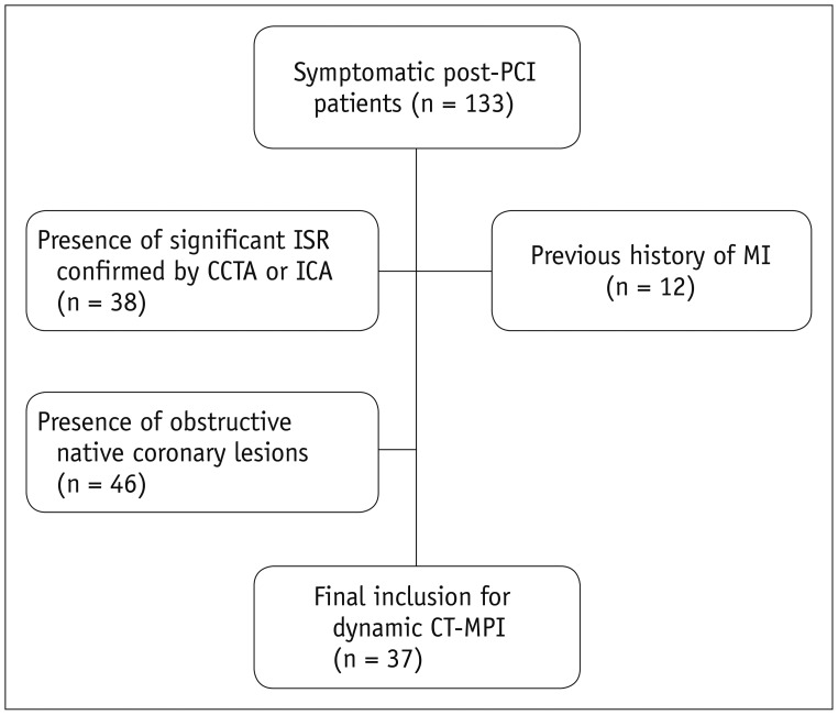
Fig. 1 Flowchart illustrating inclusion and exclusion criteria.CCTA = coronary CT angiography, CT = computed tomography, ICA = invasive coronary angiography, ISR = in-stent restenosis, MI = myocardial infarction, MPI = myocardial perfusion imaging, PCI = percutaneous coronary intervention
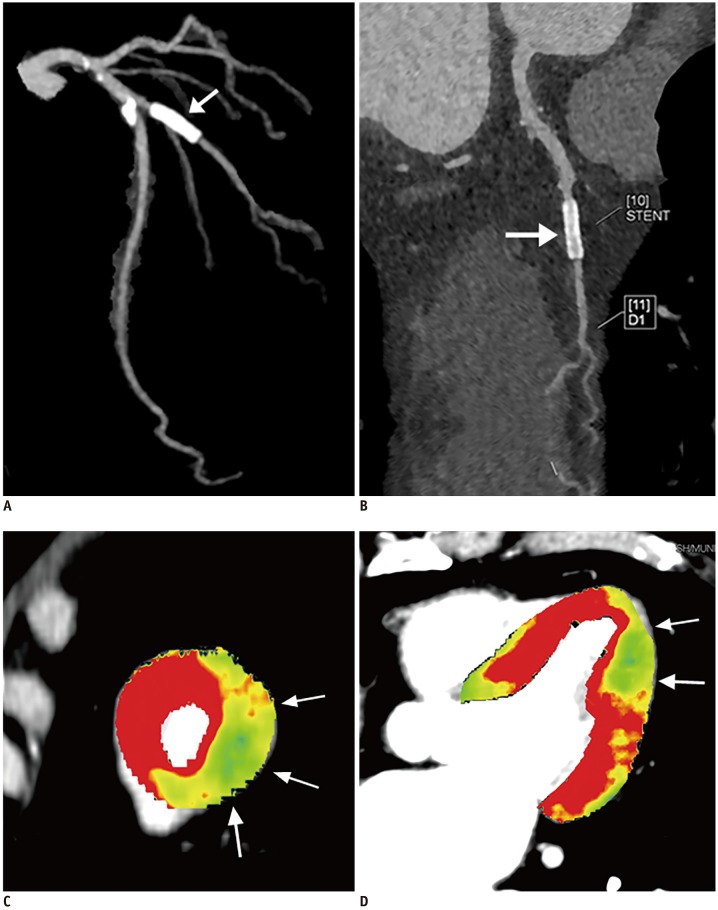
Fig. 2 Representative case of post-PCI patient with patent stent and reduced myocardial perfusion.A. 3D-MIP image showed stent implantation of first diagonal branch (arrow). B. CPR image confirmed stent patency (arrow). C. Short-axis view of MBF fusion image revealing decreased MBF in apical lateral segment (arrows), corresponding to first diagonal branch territory. D. Long-axis view of MBF fusion image demonstrating decreased MBF in apical lateral segment (arrows); mean MBF was 90.5 mL/100 mL/min. CPR = curved planar reformation, MBF = myocardial blood flow, MIP = maximum intensity projection, 3D = three-dimensional
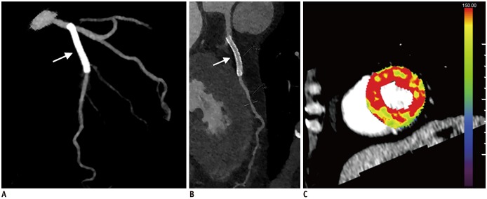
Fig. 3 Representative case of post-PCI patient with patent stent and normal myocardial perfusion.A. 3D-MIP image showing stent implantation of proximal LAD (arrow). B. CPR image confirming stent patency (arrow). C. Short-axis view of MBF fusion image revealing normal MBF throughout left ventricle. Mean MBF of LAD territory was 150.9 mL/100 mL/min. LAD = left anterior descending
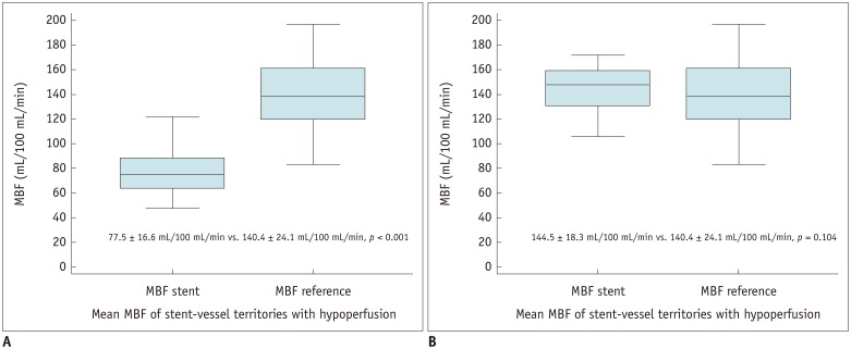
Fig. 4 Box plot of MBF for stent-vessel territories and reference territories.A. Mean MBF of stent-vessel territories with hypoperfusion was significantly lower than that of reference territories. B. Mean MBF of stent-vessel territories with hypoperfusion was similar to that of that reference territories.
Reference
-
1. Sousa JE, Serruys PW, Costa MA. New frontiers in cardiology: drug-eluting stents: part I. Circulation. 2003; 107:2274–2279. PMID: 12732594.2. Sousa JE, Serruys PW, Costa MA. New frontiers in cardiology: drug-eluting stents: part II. Circulation. 2003; 107:2383–2389. PMID: 12742968.3. Moses JW, Leon MB, Popma JJ, Fitzgerald PJ, Holmes DR, O'Shaughnessy C, et al. Sirolimus-eluting stents versus standard stents in patients with stenosis in a native coronary artery. N Engl J Med. 2003; 349:1315–1323. PMID: 14523139.
Article4. Stone GW, Ellis SG, Cox DA, Hermiller J, O'Shaughnessy C, Mann JT, et al. A polymer-based, paclitaxel-eluting stent in patients with coronary artery disease. N Engl J Med. 2004; 350:221–231. PMID: 14724301.
Article5. Dangas GD, Claessen BE, Caixeta A, Sanidas EA, Mintz GS, Mehran R. In-stent restenosis in the drug-eluting stent era. J Am Coll Cardiol. 2010; 56:1897–1907. PMID: 21109112.
Article6. Hokimoto S, Tabata N, Yamanaga K, Sueta D, Akasaka T, Tsujita K, et al. Prevalence of coronary macro- and micro-vascular dysfunctions after drug-eluting stent implantation without in-stent restenosis. Int J Cardiol. 2016; 222:185–194. PMID: 27497093.
Article7. Ong P, Athanasiadis A, Perne A, Mahrholdt H, Schäufele T, Hill S, et al. Coronary vasomotor abnormalities in patients with stable angina after successful stent implantation but without in-stent restenosis. Clin Res Cardiol. 2014; 103:11–19. PMID: 23995322.
Article8. Li Y, Yu M, Li W, Lu Z, Wei M, Zhang J. Third generation dual-source CT enables accurate diagnosis of coronary restenosis in all size stents with low radiation dose and preserved image quality. Eur Radiol. 2018; 28:2647–2654. PMID: 29349698.
Article9. Gould KL, Johnson NP, Bateman TM, Beanlands RS, Bengel FM, Bober R, et al. Anatomic versus physiologic assessment of coronary artery disease. Role of coronary flow reserve, fractional flow reserve, and positron emission tomography imaging in revascularization decision-making. J Am Coll Cardiol. 2013; 62:1639–1653. PMID: 23954338.10. El Fakhri G, Kardan A, Sitek A, Dorbala S, Abi-Hatem N, Lahoud Y, et al. Reproducibility and accuracy of quantitative myocardial blood flow assessment with (82) Rb PET: comparison with (13)N-ammonia PET. J Nucl Med. 2009; 50:1062–1071. PMID: 19525467.11. Alexánderson E, Ochoa JM, Calleja R, Juárez-Rojas JG, Prior JO, Jácome R, et al. Endothelial dysfunction in systemic lupus erythematosus: evaluation with 13N-ammonia PET. J Nucl Med. 2010; 51:1927–1931. PMID: 21078786.
Article12. Recio-Mayoral A, Rimoldi OE, Camici PG, Kaski JC. Inflammation and microvascular dysfunction in cardiac syndrome X patients without conventional risk factors for coronary artery disease. JACC Cardiovasc Imaging. 2013; 6:660–667. PMID: 23643286.
Article13. Bamberg F, Becker A, Schwarz F, Marcus RP, Greif M, von Ziegler F, et al. Detection of hemodynamically significant coronary artery stenosis: incremental diagnostic value of dynamic CT-based myocardial perfusion imaging. Radiology. 2011; 260:689–698. PMID: 21846761.
Article14. Bamberg F, Marcus RP, Becker A, Hildebrandt K, Bauner K, Schwarz F, et al. Dynamic myocardial CT perfusion imaging for evaluation of myocardial ischemia as determined by MR imaging. JACC Cardiovasc Imaging. 2014; 7:267–277. PMID: 24529887.
Article15. Coenen A, Rossi A, Lubbers MM, Kurata A, Kono AK, Chelu RG, et al. Integrating CT myocardial perfusion and CT-FFR in the work-up of coronary artery disease. JACC Cardiovasc Imaging. 2017; 10:760–770. PMID: 28109933.16. Bamberg F, Klotz E, Flohr T, Becker A, Becker CR, Schmidt B, et al. Dynamic myocardial stress perfusion imaging using fast dual-source CT with alternating table positions: initial experience. Eur Radiol. 2010; 20:1168–1173. PMID: 20333388.
Article17. Rossi A, Merkus D, Klotz E, Mollet N, de Feyter PJ, Krestin GP. Stress myocardial perfusion: imaging with multidetector CT. Radiology. 2014; 270:25–46. PMID: 24354374.
Article18. Cerqueira MD, Weissman NJ, Dilsizian V, Jacobs AK, Kaul S, Laskey WK, et al. American Heart Association Writing Group on Myocardial Segmentation and Registration for Cardiac Imaging. Standardized myocardial segmentation and nomenclature for tomographic imaging of the heart. A statement for healthcare professionals from the Cardiac Imaging Committee of the Council on Clinical Cardiology of the American Heart Association. Circulation. 2002; 105:539–542. PMID: 11815441.
Article19. Danad I, Szymonifka J, Schulman-Marcus J, Min JK. Static and dynamic assessment of myocardial perfusion by computed tomography. Eur Heart J Cardiovasc Imaging. 2016; 17:836–844. PMID: 27013250.
Article20. Kono AK, Coenen A, Lubbers M, Kurata A, Rossi A, Dharampal A, et al. Relative myocardial blood flow by dynamic computed tomographic perfusion imaging predicts hemodynamic significance of coronary stenosis better than absolute blood flow. Invest Radiol. 2014; 49:801–807. PMID: 25014013.
Article21. Monnink SH, Tio RA, Veeger NJ, Amoroso G, van Boven AJ, van Gilst WH. Exercise-induced ischemia after successful percutaneous coronary intervention is related to distal coronary endothelial dysfunction. J Investig Med. 2003; 51:221–226.
Article22. Balian V, Galli M, Marcassa C, Cecchin G, Child M, Barlocco F, et al. Intracoronary ST-segment shift soon after elective percutaneous coronary intervention accurately predicts periprocedural myocardial injury. Circulation. 2006; 114:1948–1954. PMID: 17060382.
Article23. Jaffe R, Dick A, Strauss BH. Prevention and treatment of microvascular obstruction related myocardial injury and coronary no-reflow following percutaneous coronary intervention: a systematic approach. JACC Cardiovasc Interv. 2010; 3:695–704. PMID: 20650430.24. Ikenaga H, Kurisu S, Nakao T, Kono S, Sumimoto Y, Watanabe N, et al. Predictive value of plaque morphology assessed by frequency-domain optical coherence tomography for impaired microvascular perfusion after elective stent implantation: the intracoronary electrocardiogram study. Eur Heart J Cardiovasc Imaging. 2018; 19:310–318. PMID: 28329036.
Article25. Einstein AJ, Moser KW, Thompson RC, Cerqueira MD, Henzlova MJ. Radiation dose to patients from cardiac diagnostic imaging. Circulation. 2007; 116:1290–1305. PMID: 17846343.
Article26. Zhang LJ, Wang Y, Schoepf UJ, Meinel FG, Bayer RR 2nd, Qi L, et al. Image quality, radiation dose, and diagnostic accuracy of prospectively ECG-triggered high-pitch coronary CT angiography at 70 kVp in a clinical setting: comparison with invasive coronary angiography. Eur Radiol. 2016; 26:797–806. PMID: 26382844.27. Layritz C, Schmid J, Achenbach S, Ulzheimer S, Wuest W, May M, et al. Accuracy of prospectively ECG-triggered very low-dose coronary dual-source CT angiography using iterative reconstruction for the detection of coronary artery stenosis: comparison with invasive catheterization. Eur Heart J Cardiovasc Imaging. 2014; 15:1238–1245. PMID: 24939952.
Article28. Rief M, Zimmermann E, Stenzel F, Martus P, Stangl K, Greupner J, et al. Computed tomography angiography and myocardial computed tomography perfusion in patients with coronary stents: prospective intraindividual comparison with conventional coronary angiography. J Am Coll Cardiol. 2013; 62:1476–1485. PMID: 23792193.
- Full Text Links
-
- Actions
-
Cited
- CITED
-
- Close
- Share
-
- Similar articles
-
- Dynamic CT Perfusion Imaging: State of the Art
- Stress Testing and Imaging Protocols for Myocardial Perfusion Studies
- CT Fractional Flow Reserve for the Diagnosis of Myocardial Bridging-Related Ischemia: A Study Using Dynamic CT Myocardial Perfusion Imaging as a Reference Standard
- Dynamic CT Myocardial Perfusion Imaging in Patients without Obstructive Coronary Artery Disease: Quantification of Myocardial Blood Flow according to Varied Heart Rate Increments after Stress
- Myocardial Contractility, Perfusion, and Viability Analysis Using Multidetector CT in Patients with Ischemic Heart Disease
