Computed Tomography Arthrography Findings of Idiopathic Adhesive Capsulitis of the Hip: An Analog of Adhesive Capsulitis of the Shoulder
- Affiliations
-
- 1Department of Radiology, Chung-Ang University Hospital, Seoul, Korea.
- 2Department of Orthopeadic Surgery, Chung-Ang University Hospital, Seoul, Korea. hayongch@naver.com
- KMID: 2438278
- DOI: http://doi.org/10.3348/kjr.2018.0566
Abstract
OBJECTIVE
To identify useful imaging findings for the diagnosis of idiopathic adhesive capsulitis of the hip (ACH) on computed tomography arthrography (CTA).
MATERIALS AND METHODS
Twenty-eight consecutive patients (29 hips; 7 males; mean age, 45.7 years; age range, 17-67 years) with ACH from October 2009 to March 2017 and 29 age- and sex-matched control patients from 2014 to 2016 were enrolled. All CTA images were evaluated by 2 radiologists independently for joint distensibility (anterior-posterior [AP] and superior-inferior [SI] joint cavity filling ratios), the presence of contrast filling around the ligamentum teres, and extracapsular contrast leakage. Fisher's exact test, Mann-Whitney U test, analysis of variance, and receiver operating characteristic curves were used for statistical analysis. P value less than 0.05 was considered to indicate statistical significance.
RESULTS
The anterior joint cavity was significantly more obliterated in the ACH group (mean size, 3.7-4.0 mm) than in the control group (mean size, 4.8-5.0 mm; p < 0.05). The AP filling ratio was also significantly lower in the ACH group (0.6 vs. 1.1; p < 0.05) and decreased more as the ACH stage increased (mean anterior joint cavity size: 1.15 mm in stage 3 vs. 4.68 mm in stage 1; p < 0.05). Extracapsular contrast leakage was more common in the ACH group (27-28 vs. 20-21; p = 0.041 and 0.025, respectively).
CONCLUSION
On CTA, the anterior joint cavity may have earlier and more marked obliteration than joint cavities on other sides, and may be accompanied by extracapsular contrast leakage in ACH. These CTA findings may be helpful in the diagnosis of ACH.
MeSH Terms
Figure
-
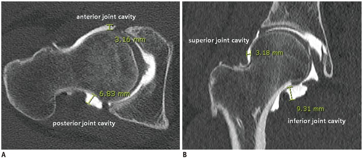
Fig. 1 Measurement of hip joint cavity.On CTA of 50-year-old male with stage 1 idiopathic ACH, widest distance of anterior joint cavity was markedly shorter (3.16 mm) than that of posterior joint cavity (6.83 mm) on mid-oblique axial image (A), resulting in low AP filling ratio (0.46). In addition, widest distance of superior joint cavity was markedly shorter (3.18 mm) than that of inferior joint cavity (9.31 mm) on mid-coronal image (B), resulting in low SI filling ratio (0.34). ACH = adhesive capsulitis of the hip, AP = anterior-posterior, CTA = computed tomography arthrography, SI = superior-inferior
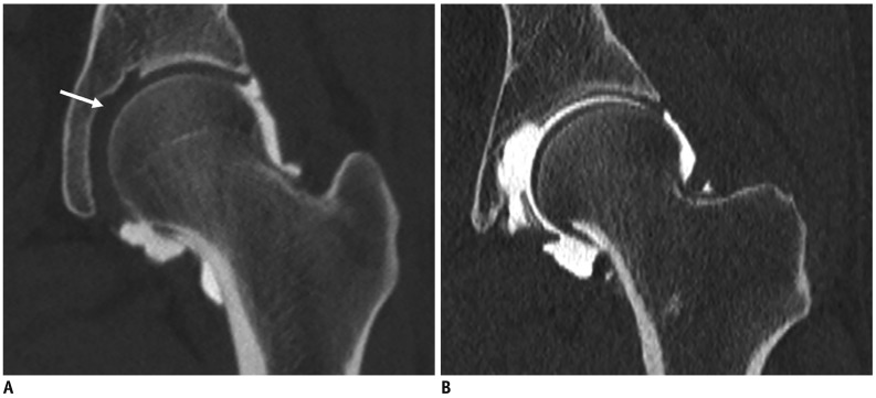
Fig. 2 Lack of contrast filling around ligamentum teres.A coronal CTA image of 40-year-old female with stage 2 idiopathic ACH showed no contrast filling along medial side of hip joint around ligamentum teres (arrow, A). However, contrast filling was normal around ligamentum teres on coronal CTA image of 40-year-old female in control group (B), which was confirmed as anterior labral tear on arthroscopy.
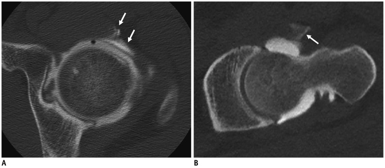
Fig. 3 Contrast leakage from hip joint capsule.Oblique axial CTA image of 37-year-old male with stage 3 idiopathic ACH showed contrast leakage from underlying anterior joint capsule (arrows, A) as well as along needle tract (arrow, B).
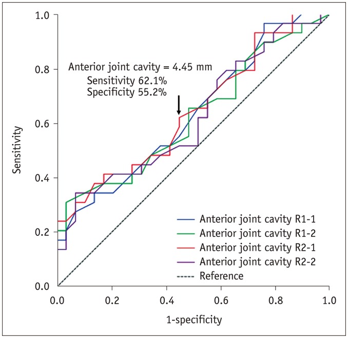
Fig. 4 Diagnostic performance of anterior hip joint cavity for diagnosis of ACH.Receiver operating characteristic curve of distance of anterior hip joint cavity showed that distance of 4.45 mm had 62.1% sensitivity and 55.2% specificity in diagnosis of ACH.
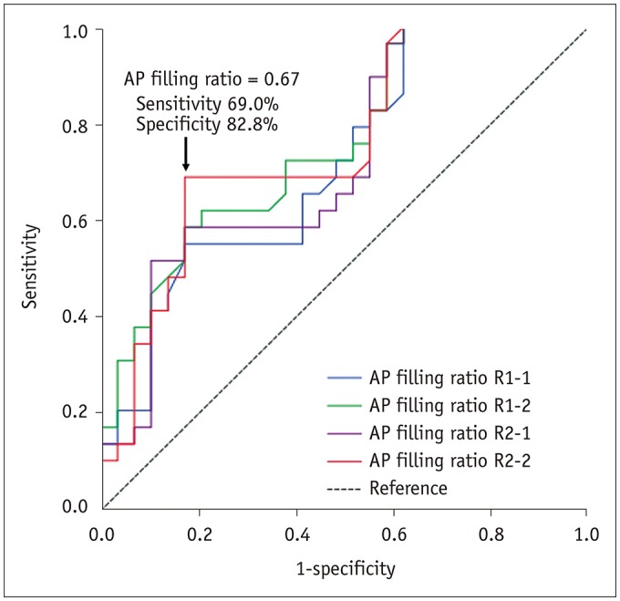
Fig. 5 Diagnostic performance of AP filling ratio for diagnosis of ACH.Receiver operating characteristic curve of AP filling ratio (anterior/posterior joint cavity) showed cut-off value of 0.67 with 69.0% sensitivity and 82.8% specificity in diagnosis of ACH.
Reference
-
1. Tamai K, Akutsu M, Yano Y. Primary frozen shoulder: brief review of pathology and imaging abnormalities. J Orthop Sci. 2014; 19:1–5. PMID: 24306579.
Article2. Hsu JE, Anakwenze OA, Warrender WJ, Abboud JA. Current review of adhesive capsulitis. J Shoulder Elbow Surg. 2011; 20:502–514. PMID: 21167743.
Article3. Connell D, Padmanabhan R, Buchbinder R. Adhesive capsulitis: role of MR imaging in differential diagnosis. Eur Radiol. 2002; 12:2100–2106. PMID: 12136330.
Article4. Emig EW, Schweitzer ME, Karasick D, Lubowitz J. Adhesive capsulitis of the shoulder: MR diagnosis. AJR Am J Roentgenol. 1995; 164:1457–1459. PMID: 7754892.
Article5. Carrillon Y, Noel E, Fantino O, Perrin-Fayolle O, Tran-Minh VA. Magnetic resonance imaging findings in idiopathic adhesive capsulitis of the shoulder. Rev Rhum Engl Ed. 1999; 66:201–206. PMID: 10339775.6. Manton GL, Schweitzer ME, Weishaupt D, Karasick D. Utility of MR arthrography in the diagnosis of adhesive capsulitis. Skeletal Radiol. 2001; 30:326–330. PMID: 11465773.
Article7. Lee MH, Ahn JM, Muhle C, Kim SH, Park JS, Kim SH, et al. Adhesive capsulitis of the shoulder: diagnosis using magnetic resonance arthrography, with arthroscopic findings as the standard. J Comput Assist Tomogr. 2003; 27:901–906. PMID: 14600458.8. Mengiardi B, Pfirrmann CW, Gerber C, Hodler J, Zanetti M. Frozen shoulder: MR arthrographic findings. Radiology. 2004; 233:486–492. PMID: 15358849.
Article9. Caroit M, Djian A, Hubault A, Normandin C, de Seze S. 2 cases of retractile capsulitis of the hip. Rev Rhum Mal Osteoartic. 1963; 30:784. PMID: 14116399.10. Looney CG, Raynor B, Lowe R. Adhesive capsulitis of the hip: a review. J Am Acad Orthop Surg. 2013; 21:749–755. PMID: 24292931.
Article11. Murphy WA, Siegel M, Gilula L. Arthrography in the diagnosis of unexplained chronic hip pain with regional osteopenia. AJR Am J Roentgenol. 1977; 129:283–287. PMID: 409164.
Article12. Lequesne M, Becker J, Bard M, Witvoet J, Postel M. Capsular constriction of the hip: arthrographic and clinical considerations. Skeletal Radiol. 1981; 6:1–10. PMID: 7466410.
Article13. Griffiths HJ, Utz R, Burke J, Bonfiglio T. Adhesive capsulitis of the hip and ankle. AJR Am J Roentgenol. 1985; 144:101–105. PMID: 3871127.
Article14. Butt SH, Muthukumar T, Cassar-Pullicino V, Mangham D. Primary synovial osteochondromatosis presenting as constrictive capsulitis. Skeletal Radiol. 2005; 34:707–713. PMID: 16132979.
Article15. Mont MA, Lindsey JM, Hungerford DS. Adhesive capsulitis of the hip. Orthopedics. 1999; 22:343–345. PMID: 10192266.
Article16. Byrd JW, Jones KS. Adhesive capsulitis of the hip. Arthroscopy. 2006; 22:89–94. PMID: 16399467.
Article17. Hulstyn MJ, Weiss AP. Adhesive capsulitis of the shoulder. Orthop Rev. 1993; 22:425–433. PMID: 8479787.18. Uitvlugt G, Detrisac DA, Johnson LL, Austin MD, Johnson C. Arthroscopic observations before and after manipulation of frozen shoulder. Arthroscopy. 1993; 9:181–185. PMID: 8461078.
Article19. Neviaser RJ, Neviaser TJ. The frozen shoulder diagnosis and management. Clin Orthop Relat Res. 1987; (223):59–64.
Article20. Hannafin J, DiCarlo E, Wickiewicz T, Warren R. Adhesive capsulitis: capsular fibroplasia of the glenohumeral joint. J Shoulder Elbow Surg. 1994; 3:435.21. Neviaser AS, Hannafin JA. Adhesive capsulitis: a review of current treatment. Am J Sports Med. 2010; 38:2346–2356. PMID: 20110457.
- Full Text Links
-
- Actions
-
Cited
- CITED
-
- Close
- Share
-
- Similar articles
-
- Adhesive Capsulitis of the Shoulder
- The Effects of Stellate Ganglion Block in Adhesive Capsulitis of the Shoulder
- The Comparison of Therapeutic Effect of Shoulder Pain with Simple and Mixed Type in Patients with Adhesive Capsulitis
- Ultrasonography in Adhesive Capsulitis of Shoulder
- Ultrasonography and arthrography in rotator cuff lesions: algorithmic approach
