Optimal Kiloelectron Volt for Noise-Optimized Virtual Monoenergetic Images of Dual-Energy Pediatric Abdominopelvic Computed Tomography: Preliminary Results
- Affiliations
-
- 1Department of Radiology, Seoul National University Hospital, Seoul, Korea. iater@snu.ac.kr
- 2Department of Radiology, Seoul National University College of Medicine, Seoul, Korea.
- 3Institute of Radiation Medicine, Seoul National University Medical Research Center, Seoul, Korea.
- 4Department of Radiology, Kyung Hee University Hospital, Seoul, Korea.
- 5Department of Radiology, Seoul Metropolitan Government-Seoul National University Boramae Medical Center, Seoul, Korea.
- 6Siemens Healthineers, Seoul, Korea.
- 7Siemens Healthineers, Forchheim, Germany.
- KMID: 2438259
- DOI: http://doi.org/10.3348/kjr.2017.0507
Abstract
OBJECTIVE
To compare quantitative and qualitative image quality parameters in pediatric abdominopelvic dual-energy CT (DECT) using noise-optimized virtual monoenergetic image (VMI) and conventional VMI at different kiloelectron volt (keV) levels.
MATERIALS AND METHODS
Thirty-six consecutive abdominopelvic DECT scans were retrospectively included. Noise-optimized VMI and conventional VMI were reconstructed at seven energy levels, from 40 keV to 100 keV at 10 keV intervals. The contrast-to-noise ratio (CNR) and signal-to-noise ratio (SNR) of the liver, pancreas, and aorta were objectively measured and compared. Image quality was evaluated subjectively regarding image noise, image blurring of solid organ, bowel image quality and severity of beam-hardening artifacts. Optimal monoenergetic levels in keV for both algorithms were determined based on overall image quality score.
RESULTS
The maximal CNR and SNR values for all investigated organs were observed at 40 keV in noise-optimized VMI (CNR and SNR of liver, pancreas, aorta in order [CNR; 20.93, 17.34, 46.75: SNR; 37.39, 33.80, 63.21]), at 60-70 keV and at 70 keV in conventional VMI (CNR; 8.12, 5.67, 15.97: SNR; 19.57, 16.66, 26.65). In qualitative image analysis, noise-optimized VMI and conventional VMI showed the best overall image quality scores at 60 keV and at 70 keV, respectively. Noise-optimized VMI at 60 keV showed superior CNRs, SNRs, and overall image quality scores compared to conventional VMI at 70 keV (p < 0.001).
CONCLUSION
Optimal energy levels for noise-optimized VMI and conventional VMI were 60 keV and at 70 keV, respectively. Noise-optimized VMI shows superior CNRs, SNRs and subjective image quality over conventional VMI, at the optimal energy level.
Keyword
Figure
-
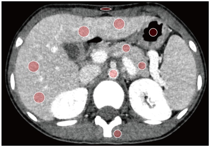
Fig. 1 Manually drawn regions of interest in liver, pancreas, aorta, paraspinal muscle, subcutaneous fat of anterior abdominal wall, and air column.All measurements were kept constant across VMI levels by using copy-and-paste function at workstation. VMI = virtual monoenergetic image
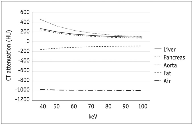
Fig. 2 Graphs of mean attenuation of liver, pancreas, aorta, subcutaneous fat, and air in mean of noise-optimized and conventional algorithms.HU = Hounsfield unit, keV = kiloelectron volt
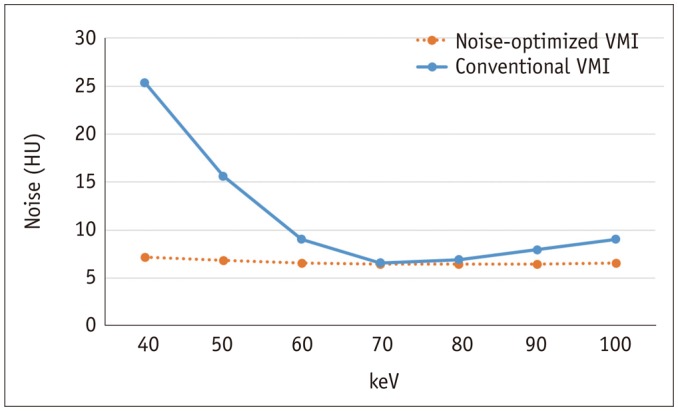
Fig. 3 Graphs of image noise in noise-optimized and conventional algorithms.
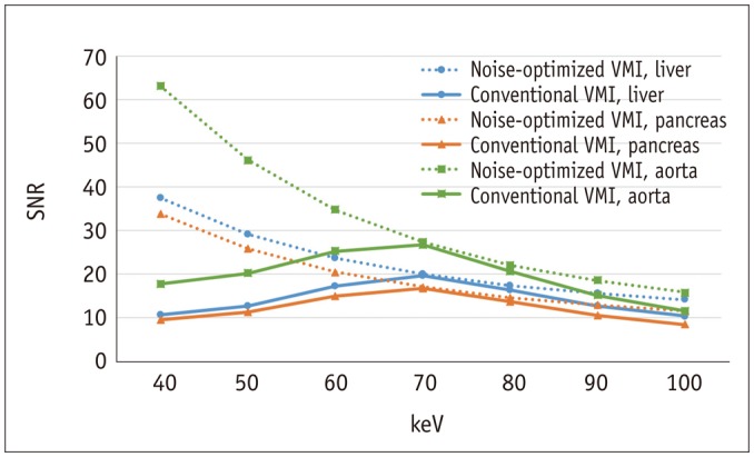
Fig. 4 Graphs showing SNR values of liver, pancreas, and aorta in noise-optimized and conventional VMIs.SNR was significantly higher in noise-optimized algorithm compared to conventional algorithm at all VMI energy levels. SNR = signal-to-noise ratio
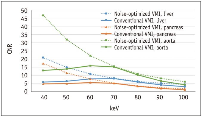
Fig. 5 Graphs of CNR values of liver, pancreas, and aorta in noise-optimized and conventional VMIs.CNR was significantly higher in noise-optimized algorithm compared to conventional algorithm in all VMI energy levels, except for 70 keV. CNR = contrast-to-noise ratio
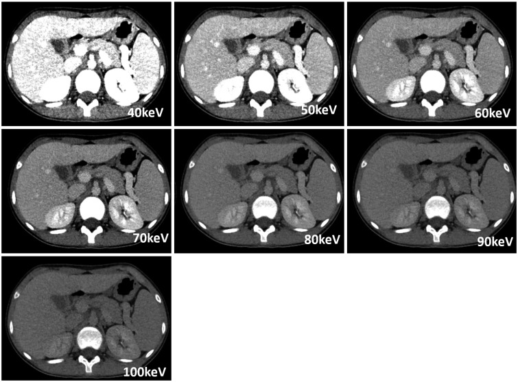
Fig. 6 Images obtained by noise-optimized algorithm with 40–100 keV energy levels.Best CNR and SNR were obtained at 40 keV. In subjective analysis, 60 keV scored best in terms of overall image quality. Taken together, 60 keV was considered to be optimal VMI energy level for noise-optimized algorithm.
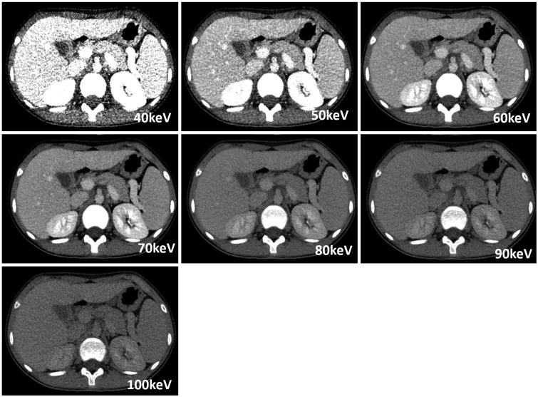
Fig. 7 Images obtained by conventional algorithm with 40–100 keV energy levels.Best CNR was obtained at 60–70 keV, and best SNR was obtained at 70 keV. Further, 70 keV achieved best overall image quality score in subjective image analysis. Therefore, 70 keV was considered to be optimal VMI energy level for conventional algorithm.
Reference
-
1. Goo HW, Goo JM. Dual-energy CT: new horizon in medical imaging. Korean J Radiol. 2017; 18:555–569. PMID: 28670151.
Article2. Yu L, Leng S, McCollough CH. Dual-energy CT-based monochromatic imaging. AJR Am J Roentgenol. 2012; 199(5 Suppl):S9–S15. PMID: 23097173.
Article3. Apfaltrer P, Sudarski S, Schneider D, Nance JW Jr, Haubenreisser H, Fink C, et al. Value of monoenergetic low-kV dual energy CT datasets for improved image quality of CT pulmonary angiography. Eur J Radiol. 2014; 83:322–328. PMID: 24361061.
Article4. Schneider D, Apfaltrer P, Sudarski S, Nance JW Jr, Haubenreisser H, Fink C, et al. Optimization of kiloelectron volt settings in cerebral and cervical dual-energy CT angiography determined with virtual monoenergetic imaging. Acad Radiol. 2014; 21:431–436. PMID: 24594412.
Article5. Bamberg F, Dierks A, Nikolaou K, Reiser MF, Becker CR, Johnson TR. Metal artifact reduction by dual energy computed tomography using monoenergetic extrapolation. Eur Radiol. 2011; 21:1424–1429. PMID: 21249370.
Article6. Leschka S, Stolzmann P, Schmid FT, Scheffel H, Stinn B, Marincek B, et al. Low kilovoltage cardiac dual-source CT: attenuation, noise, and radiation dose. Eur Radiol. 2008; 18:1809–1817. PMID: 18392829.
Article7. Grant KL, Flohr TG, Krauss B, Sedlmair M, Thomas C, Schmidt B. Assessment of an advanced image-based technique to calculate virtual monoenergetic computed tomographic images from a dual-energy examination to improve contrast-to-noise ratio in examinations using iodinated contrast media. Invest Radiol. 2014; 49:586–592. PMID: 24710203.
Article8. Albrecht MH, Scholtz JE, Kraft J, Bauer RW, Kaup M, Dewes P, et al. Assessment of an advanced monoenergetic reconstruction technique in dual-energy computed tomography of head and neck cancer. Eur Radiol. 2015; 25:2493–2501. PMID: 25680727.
Article9. Meier A, Wurnig M, Desbiolles L, Leschka S, Frauenfelder T, Alkadhi H. Advanced virtual monoenergetic images: improving the contrast of dual-energy CT pulmonary angiography. Clin Radiol. 2015; 70:1244–1251. PMID: 26231468.
Article10. Albrecht MH, Scholtz JE, Hüsers K, Beeres M, Bucher AM, Kaup M, et al. Advanced image-based virtual monoenergetic dual-energy CT angiography of the abdomen: optimization of kiloelectron volt settings to improve image contrast. Eur Radiol. 2016; 26:1863–1870. PMID: 26334508.
Article11. Zhu X, McCullough WP, Mecca P, Servaes S, Darge K. Dual-energy compared to single-energy CT in pediatric imaging: a phantom study for DECT clinical guidance. Pediatr Radiol. 2016; 46:1671–1679. PMID: 27518078.
Article12. Siegel MJ, Curtis WA, Ramirez-Giraldo JC. Effects of dual-energy technique on radiation exposure and image quality in pediatric body CT. AJR Am J Roentgenol. 2016; 207:826–835. PMID: 27490819.
Article13. Nievelstein RA, van Dam IM, van der Molen AJ. Multidetector CT in children: current concepts and dose reduction strategies. Pediatr Radiol. 2010; 40:1324–1344. PMID: 20535463.
Article14. Lubner MG, Pickhardt PJ, Tang J, Chen GH. Reduced image noise at low-dose multidetector CT of the abdomen with prior image constrained compressed sensing algorithm. Radiology. 2011; 260:248–256. PMID: 21436086.
Article15. Marin D, Nelson RC, Schindera ST, Richard S, Youngblood RS, Yoshizumi TT, et al. Low-tube-voltage, high-tube-current multidetector abdominal CT: improved image quality and decreased radiation dose with adaptive statistical iterative reconstruction algorithm--initial clinical experience. Radiology. 2010; 254:145–153. PMID: 20032149.
Article16. Winklhofer S, Lambert JW, Sun Y, Wang ZJ, Sun DS, Yeh BM. Pelvic beam-hardening artifacts in dual-energy CT image reconstructions: occurrence and impact on image quality. AJR Am J Roentgenol. 2017; 208:114–123. PMID: 27786561.
Article17. MacDougall RD, Kleinman PL, Yu L, Lee EY. Pediatric thoracic CT angiography at 70 kV: a phantom study to investigate the effects on image quality and radiation dose. Pediatr Radiol. 2016; 46:1114–1119. PMID: 26987734.18. Yu L, Bruesewitz MR, Thomas KB, Fletcher JG, Kofler JM, McCollough CH. Optimal tube potential for radiation dose reduction in pediatric CT: principles, clinical implementations, and pitfalls. Radiographics. 2011; 31:835–848. PMID: 21571660.
Article19. Verdun FR, Lepori D, Monnin P, Valley JF, Schnyder P, Gudinchet F. Management of patient dose and image noise in routine pediatric CT abdominal examinations. Eur Radiol. 2004; 14:835–841. PMID: 14722730.
Article20. Sudarski S, Apfaltrer P, Nance JW Jr, Schneider D, Meyer M, Schoenberg SO, et al. Optimization of keV-settings in abdominal and lower extremity dual-source dual-energy CT angiography determined with virtual monoenergetic imaging. Eur J Radiol. 2013; 82:e574–e581. PMID: 23763858.
Article21. Pomerantz SR, Kamalian S, Zhang D, Gupta R, Rapalino O, Sahani DV, et al. Virtual monochromatic reconstruction of dual-energy unenhanced head CT at 65-75 keV maximizes image quality compared with conventional polychromatic CT. Radiology. 2013; 266:318–325. PMID: 23074259.
Article22. Matsumoto K, Jinzaki M, Tanami Y, Ueno A, Yamada M, Kuribayashi S. Virtual monochromatic spectral imaging with fast kilovoltage switching: improved image quality as compared with that obtained with conventional 120-kVp CT. Radiology. 2011; 259:257–262. PMID: 21330561.
Article
- Full Text Links
-
- Actions
-
Cited
- CITED
-
- Close
- Share
-
- Similar articles
-
- Imaging Findings of Peripheral Arterial Disease on Lower-Extremity CT Angiography Using a Virtual Monoenergetic Imaging Algorithm
- Subjective and Objective Assessment of Monoenergetic and Polyenergetic Images Acquired by Dual-Energy CT in Breast Cancer
- Virtual Monochromatic Image Quality from Dual-Layer Dual-Energy Computed Tomography for Detecting Brain Tumors
- Dual-Energy Computed Tomography Arthrography of the Shoulder Joint Using Virtual Monochromatic Spectral Imaging: Optimal Dose of Contrast Agent and Monochromatic Energy Level
- Dual-Energy CT: New Horizon in Medical Imaging
