J Korean Ophthalmol Soc.
2019 Feb;60(2):201-204. 10.3341/jkos.2019.60.2.201.
Homonymous Quadrantanopia Caused by Occipital Lobe Ulegyria
- Affiliations
-
- 1Department of Ophthalmology, Yeungnam University College of Medicine, Daegu, Korea. eyekwj@ynu.ac.kr
- KMID: 2438082
- DOI: http://doi.org/10.3341/jkos.2019.60.2.201
Abstract
- PURPOSE
We report a case of homonymous quadrantanopia caused by occipital lobe ulegyria.
CASE SUMMARY
A 23-year-female was referred to our clinic because of a visual field defect incidentally discovered during preoperative evaluation for refractive surgery at another clinic. However, she did not report any symptoms. She had no systemic diseases. Visual acuity was 20/20 in both eyes, and the color vision test was normal. Both pupils exhibited normal responses to light and near stimulations. In fundus examinations, the right optic disc was normal and the left contained drusen. Automated perimetry revealed right lower homonymous quadrantanopia with macular sparing. Brain magnetic resonance imaging revealed areas of ulegyria involving the left occipital lobe, consistent with the visual field defect. A follow-up visual field test performed 5 months later yielded the same result.
CONCLUSIONS
Neuroimaging should be performed in patients with homonymous visual field defects to determine the location and etiology of the brain lesions. Occipital lobe ulegyria can cause homonymous quadrantanopia in the absence of any neurological problem.
Keyword
MeSH Terms
Figure
-
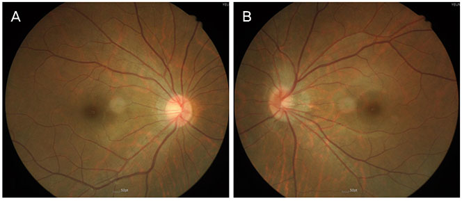
Figure 1 Images of the fundus photograph at the initial visit. The right eye showed normal appearance of optic disc (A). The left eye showed optic disc drusen (B).
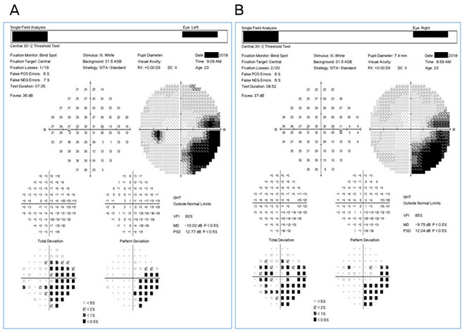
Figure 2 The result of visual field test (Humphrey Field Analyzer, Carl Zeiss Meditec, Inc., Dublin, OH, USA) performed at the initial visit. The visual field test demonstrated the right lower homonymous quadrantanopia with macular sparing (A: left eye, B: right eye).
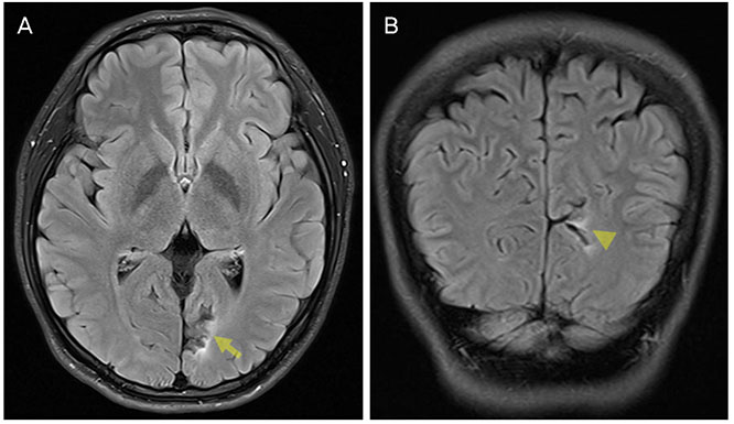
Figure 3 The coronal and axial view of the brain magnetic resonance imaging (MRI, A: axial view, B: coronal view). The MRI demonstrates the focal involvement of ulegyria at the left occipital lobe (A: arrow, B: arrowhead).
Reference
-
1. Miller NR, Subramanian PS, Patel VR. Walsh and Hoyt's clinical neuro-ophthalmology: the essentials. 3rd ed. Philadelphia: Lippincott Williams & Wilkins;2016. p. 209–241.2. Fraser JA, Newman NJ, Biousse V. Disorders of the optic tract, radiation, and occipital lobe. Handb Clin Neurol. 2011; 102:205–221.
Article3. Ogawa K, Ishikawa H, Suzuki Y, et al. Clinical study of the visual field defects caused by occipital lobe lesions. Cerebrovasc Dis. 2014; 37:102–108.
Article4. Lee HJ, Maeng YH, Jeong J, et al. Occipital lobe metastasis of hepatocellular carcinoma presenting as homonymous hemianopia. J Korean Ophthalmol Soc. 2017; 58:488–492.
Article5. Liu G, Volpe N, Galetta S. Liu, Volpe, and Galetta's neuro-ophthalmology. 3rd ed. Philadelphia: Elsevier;2019. p. 293–339.6. Pane A, Miller N, Burdon M. The neuro-ophthalmology survival guide. 2nd ed. Beijing: Elsevier;2017. p. 92–98.7. Gil-Nagel A, García Morales I, Jiménez Huete A, et al. Occipital lobe epilepsy secondary to ulegyria. J Neurol. 2005; 252:1178–1185.
Article8. Kim HW, Cho KY, Lee MC, et al. A study of ulegyria as pathognomonic aspects of congenital bilateral perisylvian syndrome. J Korean Neurosurg Soc. 2005; 37:124–128.9. Nikas I, Dermentzoglou V, Theofanopoulou M, Theodoropoulos V. Parasagittal lesions and ulegyria in hypoxic-ischemic encephalopathy: neuroimaging findings and review of the pathogenesis. J Child Neurol. 2008; 23:51–58.
Article
- Full Text Links
-
- Actions
-
Cited
- CITED
-
- Close
- Share
-
- Similar articles
-
- Neurocysticercosis Presenting as Homonymous Hemianopia
- Bilateral Occipital Lobe Infarction Presenting as Bilateral Inferior Altitudinal Defects
- Occipital Lobe Epilepsy with Hemicrania Epileptica
- Occipital Lobe Metastasis of Hepatocellular Carcinoma Presenting as Homonymous Hemianopia
- Reversible Homonymous Hemianopia Associated with Focal Hyperperfusion in Hyperglycemic State
