J Korean Ophthalmol Soc.
2017 Apr;58(4):488-492. 10.3341/jkos.2017.58.4.488.
Occipital Lobe Metastasis of Hepatocellular Carcinoma Presenting as Homonymous Hemianopia
- Affiliations
-
- 1Department of Ophthalmology, Jeju National University School of Medicine, Jeju, Korea. ilovelhj@hanmail.net
- 2Department of Pathology, Jeju National University School of Medicine, Jeju, Korea.
- 3Department of Neurosurgery, Jeju National University School of Medicine, Jeju, Korea.
- 4Department of Internal Medicine, Jeju National University School of Medicine, Jeju, Korea.
- KMID: 2376609
- DOI: http://doi.org/10.3341/jkos.2017.58.4.488
Abstract
- PURPOSE
To report brain metastasis of hepatocellular carcinoma presenting as homonymous hemianopia.
CASE SUMMARY
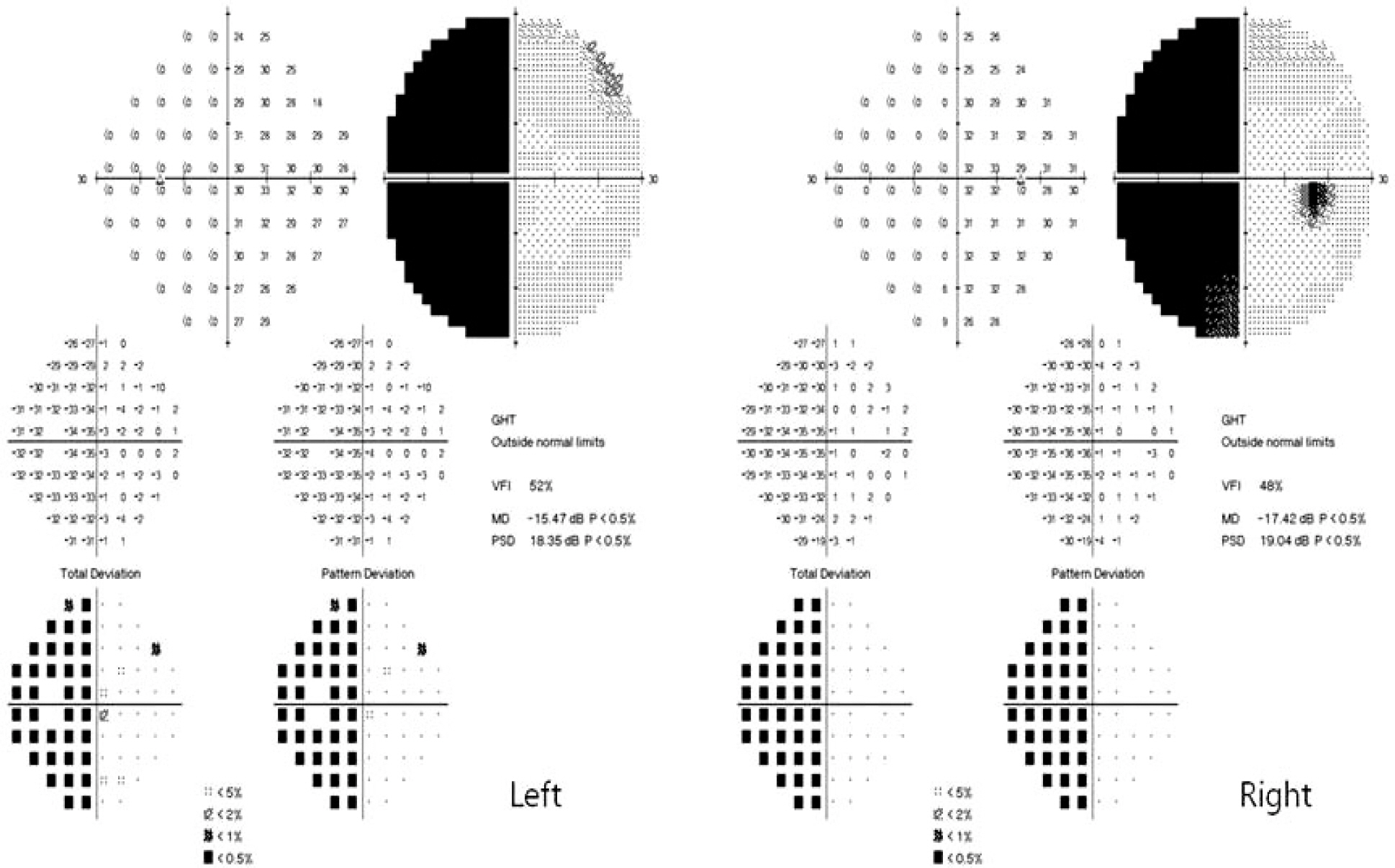
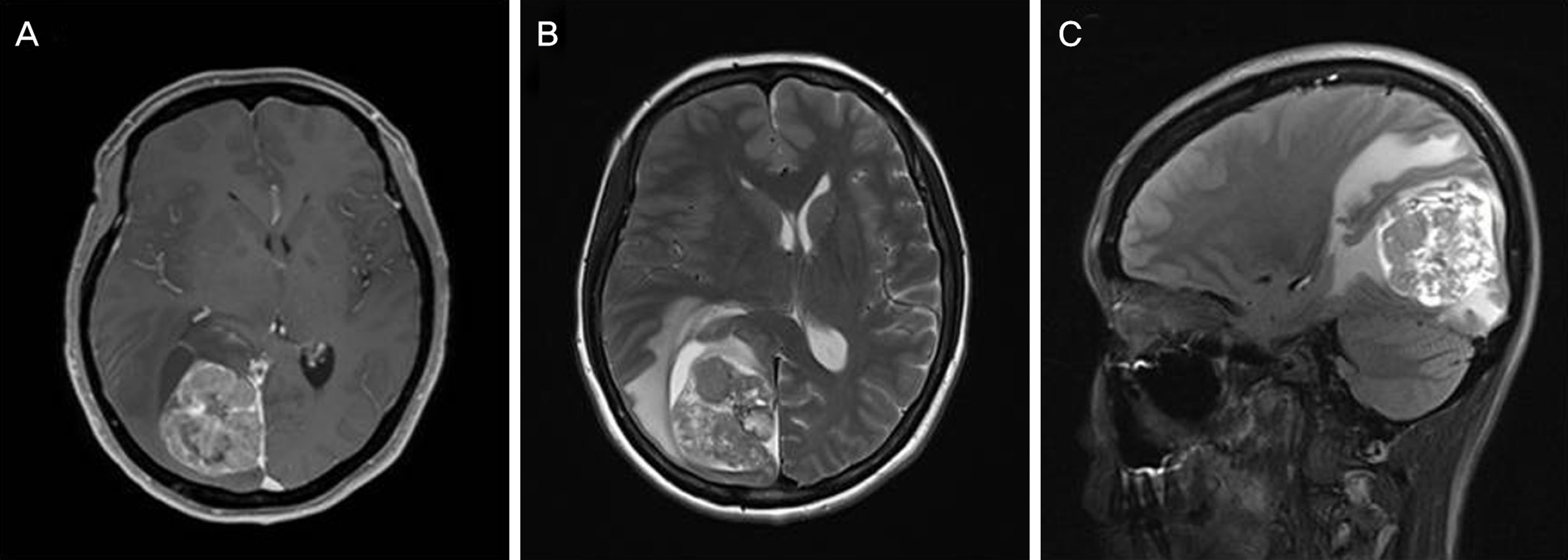
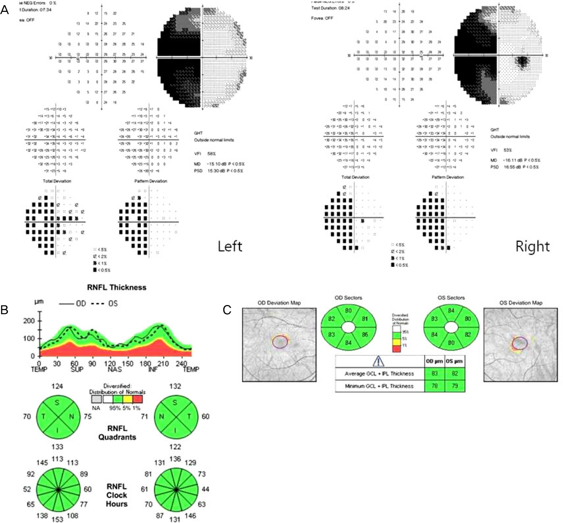
A 51-year-old female with a history of hepatectomy and diagnosis of hepatocellular carcinoma (HCC) 19 months earlier was referred to our neuro-ophthalmology clinic for evaluation due to headache and decreased visual acuity over the past several months. Best visual acuity was 20/20, and the results of all other aspects of our examination were normal except Humphrey automatic perimetry, which showed complete left homonymous hemianopia. Brain magnetic resonance imaging showed a large mass in the right occipital lobe. Craniotomy and removal of tumor were performed. HCC was confirmed by histopathologic examination.
CONCLUSIONS
Metastasis of hepatocellular carcinoma to the occipital lobe is extremely rare but can present as homonymous hemianopia. Therefore, clinicians should be aware of this when examining a patient with a history of HCC.
Keyword
MeSH Terms
Figure
Cited by 1 articles
-
Homonymous Quadrantanopia Caused by Occipital Lobe Ulegyria
Junyeop Lee, Won Jae Kim
J Korean Ophthalmol Soc. 2019;60(2):201-204. doi: 10.3341/jkos.2019.60.2.201.
Reference
-
References
1. Oh CM, Won YJ, Jung KW. . Cancer statistics in Korea: in-cidence, mortality, survival, and prevalence in 2013. Cancer Res Treat. 2016; 48:436–50.
Article2. Tsai JF, Jeng JE, Ho MS. . Effect of hepatitis C and B virus in-fection on risk of hepatocellular carcinoma: a prospective study. Br J Cancer. 1997; 76:968–74.
Article3. Choi HJ, Cho BC, Sohn JH. . Brain metastases from hep-atocellular carcinoma: prognostic factors and outcome: brain meta-stasis from HCC. J Neurooncol. 2009; 91:307–13.4. Jiang XB, Ke C, Zhang GH. . Brain metastases from hep-atocellular carcinoma: clinical features and prognostic factors. BMC Cancer. 2012; 12:49.
Article5. Hsu SY, Chang FL, Sheu MM, Tsai RK. . Homonymous hemianopia caused by solitary skull metastasis of hepatocellular carcinoma. J Neuroophthalmol. 2008; 28:51–4.
Article6. Shim YS, Ahn JY, Cho JH, Lee KS. . Solitary skull metastasis as ini-tial manifestation of hepatocellular carcinoma. World J Surg Oncol. 2008; 6:66.
Article7. Seinfeld J, Wagner AS, Kleinschmidt-DeMasters BK. . Brain meta-stases from hepatocellular carcinoma in US patients. J Neurooncol. 2006; 76:93–8.
Article8. Park TY, Na YC, Lee WH. . Treatment options of metastatic brain tumors from hepatocellular carcinoma: surgical resection vs. gamma knife radiosurgery vs. whole brain radiation therapy. Brain Tumor Res Treat. 2013; 1:78–84.9. Shinoura N, Suzuki Y, Yamada R. . Relationships between brain tumor and optic tract or calcarine fissure are involved in visu-al field deficits after surgery for brain tumor. Acta Neurochir (Wien). 2010; 152:637–42.
Article
- Full Text Links
- Actions
-
Cited
- CITED
-
- Close
- Share
- Similar articles
-
- Neurocysticercosis Presenting as Homonymous Hemianopia
- Reversible Homonymous Hemianopia Associated with Focal Hyperperfusion in Hyperglycemic State
- A Case of Multiple Sclerosis in Child Showing Homonymous Hemianopia
- Correlation of OCT and Hemifield Pattern VEP in Hemianopia
- Posterior Type of Alzheimer's Dementia Presenting with Homonymous Hemianopsia


