Prosthetic rehabilitation for patient with hemi-maxillectomy: Obturator combined with a hybrid telescopic double crown using friction pin
- Affiliations
-
- 1Department of Prosthodontics, School of Dentistry, Kyungpook National University, Daegu, Republic of Korea. prosth95@knu.ac.kr
- KMID: 2432368
- DOI: http://doi.org/10.14368/jdras.2018.34.4.317
Abstract
- When oral cancer develops in the maxilla, oro-nasal communication occurs after surgical treatment including removal of the primary site. Restoration through an obturator is necessary to prevent food from storing due to non-oral opening, and to ensure proper pronunciation and aesthetic restoration. In this case, the patient was treated with right hemi-maxillectomy due to oral cancer and has residual abutment and poor periodontal support due to the effect of head and neck radiotherapy. The obturator was treated with a hybrid telescopic double crown denture. Reporting a successful prognosis in 18 months of follow-up.
MeSH Terms
Figure
-
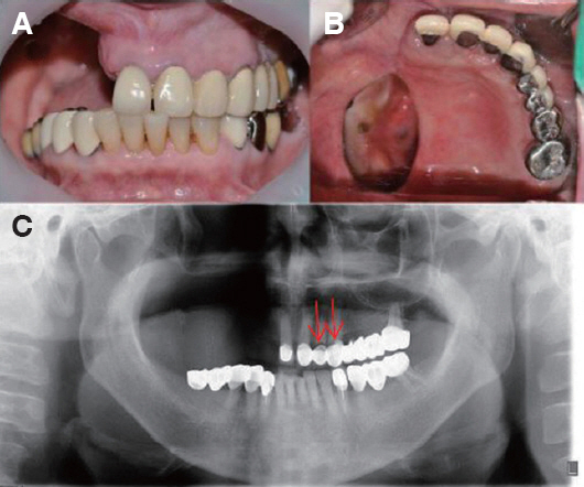
Fig. 1 Initial intraoral photograph and panoramic radiograph. (A) Frontal view, (B) Occlusal view, (C) Panoramic radiograph. Arrow: cervical caries on #22, 23.
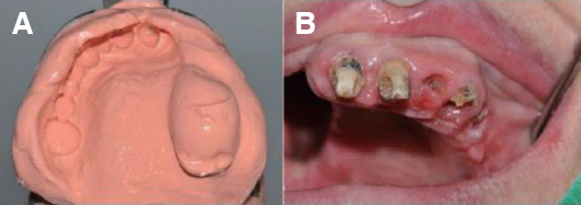
Fig. 2 Pre-prosthodontic treatment of abutments. (A) Maxillary alginate impression for individual tray, (B) Caries were removed.
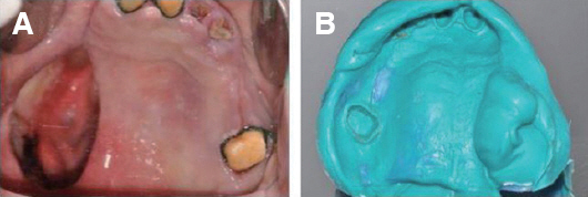
Fig. 3 Impression of maxillary abutments. (A) Preparation of abutments, (B) Impression taking for inner crowns.
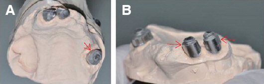
Fig. 4 Inner crowns of double crown denture (A) Occlusal view, (B) Inner crown of #11, 21 abutments. Arrow: space for friction pin.
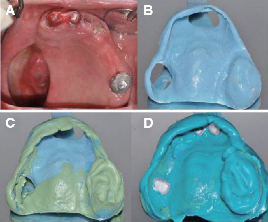
Fig. 5 Maxillary impression procedure. (A) Inner crowns were splinted, (B) Open tray modification, (C) After onestep border molding, (D) Pick up impression of inner crown.
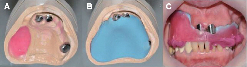
Fig. 6 VD/CR recording. (A) Maxillary master cast, (B) Recording base fabrication, (C) CR Bite registration.
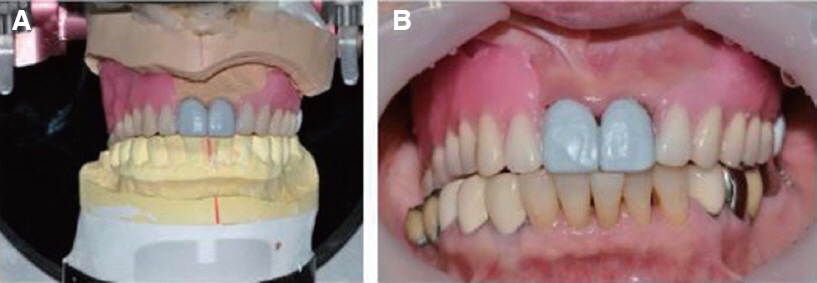
Fig. 7 Wax denture. (A) Wax denture in Articulator, (B) Wax denture try in.
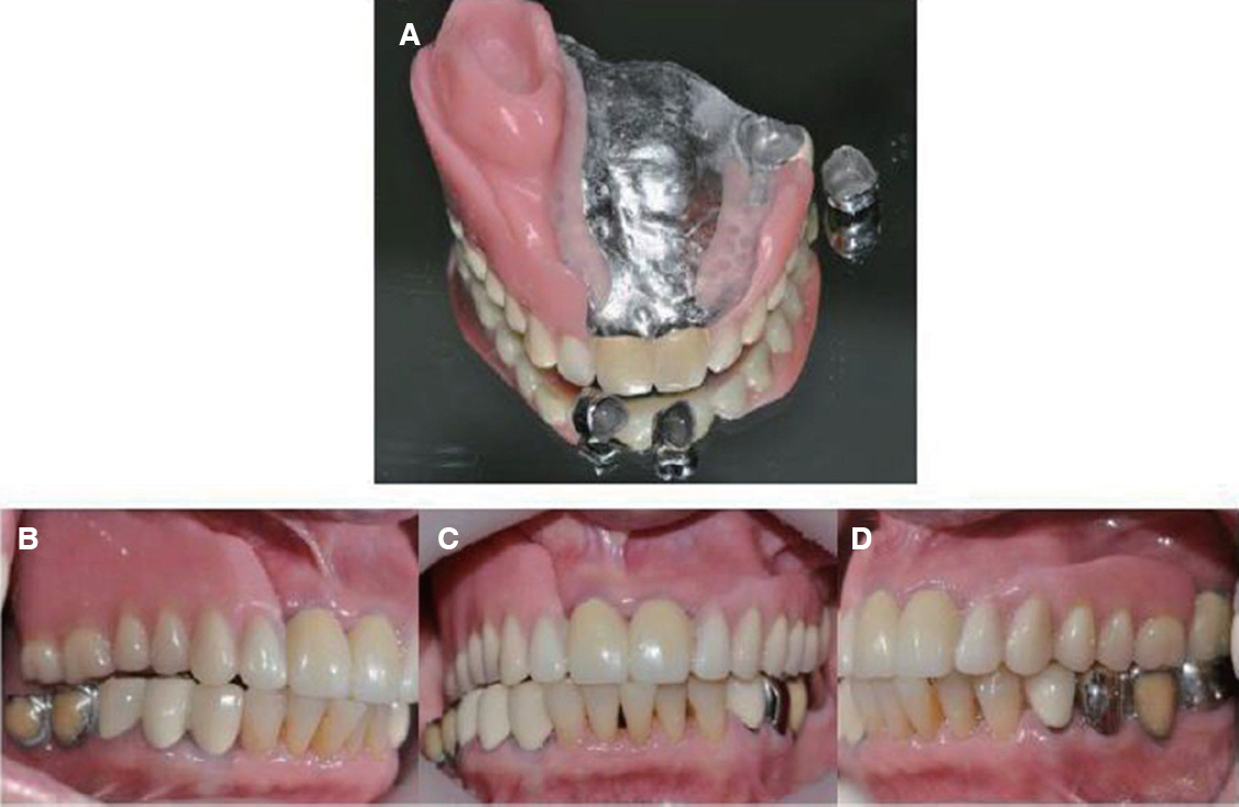
Fig. 8 Definitive prosthesis. (A) Inner crown and double crown RPD, (B) Right buccal view, (C) Frontal view, (D) Left buccal view.
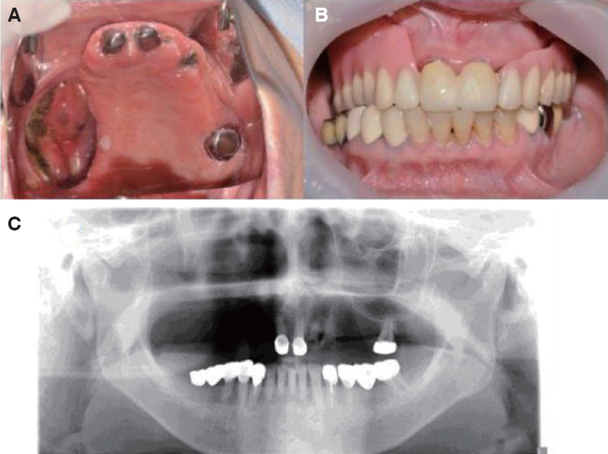
Fig. 9 18 month follow up check after delivery. (A) Occlusal view of inner crown, (B) Frontal view with denture seated, (C) Panoramic radiograph.
Reference
-
References
1. National Cancer Information Center. Available from: https://www.cancer.go.kr/lay1/program/S1T211C223/cancer/view.do?cancer_seq=3461&menu_seq=3466 (updated 2018 Aug 15).2. Rogers SN, Lowe D, McNally D, Brown JS, Vaughan ED. Health-related quality of life of after maxillectomy: a comparison between prosthetic obturation and free flap. J Oral Maxillofac Surg. 2003; 61:174–81. DOI: 10.1053/joms.2003.50044. PMID: 12618993.3. Minsley GE, Warren DW, Hinton V. Physiologic responses to maxillary resection and subsequent obturation. J Prosthet Dent. 1987; 57:338–44. DOI: 10.1016/0022-3913(87)90309-X. PMID: 3471944.4. Devlin H, Barker GR. Prosthetic rehabilitation of the edentulous patient requiring a partial maxillectomy. J Prosthet Dent. 1992; 67:223–7. DOI: 10.1016/0022-3913(92)90458-M. PMID: 1538331.5. Keyf F. Obturator prostheses for hemimaxillectomy patients. J Oral Rehabil. 2001; 28:821–9. DOI: 10.1111/j.1365-2842.2001.00754.x. PMID: 11580820.6. Weber H, Frank G. Spark erosion procedure: a method for extensive combined fixed and removable prosthodontic care. J Prosthet Dent. 1993; 69:222–7. DOI: 10.1016/0022-3913(93)90144-D. PMID: 8094098.7. Aramany MA. Basic principles of obturator design for partially edentulous patients. Part I: classification. J Prosthet Dent. 1978; 40:554–7. DOI: 10.1016/0022-3913(78)90092-6. PMID: 364015.8. Aramany MA. Basic principles of obturator design for partially edentulous patients. Part II: Design principles. J Prosthet Dent. 1978; 40:656–62. DOI: 10.1016/0022-3913(78)90065-3. PMID: 364026.9. Breitman JB, Nakamura S, Freedman AL, Yalisove IL. Telescopic retainers: an old or new solution? A second chance to have normal dental function. J Prosthodont. 2012; 21:79–83. DOI: 10.1111/j.1532-849X.2011.00797.x. PMID: 22126322.10. Wenz HJ, Hertrampf K, Lehmann KM. Clinical longevity of removable partial dentures retained by telescopic crowns: outcome of the double crown with clearance fit. Int J Prosthodont. 2001; 14:207–13. PMID: 11484566.11. Widbom T, Löfquist L, Widbom C, Söderfeldt B, Kronström M. Tooth-supported telescopic crown-retained dentures: an up to 9-year retrospective clinical follow-up study. Int J Prosthodont. 2004; 17:29–34. PMID: 15008229.12. Ishida K, Nogawa T, Takayama Y, Saito M, Yokoyama A. Prognosis of double crown-retained removable dental prostheses compared with claspretained removable dental prostheses: A retrospective study. J Prosthodont Res. 2017; 61:268–275. DOI: 10.1016/j.jpor.2016.12.006. PMID: 28073636.13. Sethuram AK, Sahoo N, Sandhu H, Radhakrishnan V. Rehabilitation of a maxillectomy case with telescopic crowns: a case report. J Indian Prosthodont Soc. 2013; 13:236–9. DOI: 10.1007/s13191-012-0147-4. PMID: 24431740. PMCID: PMC3732727.
- Full Text Links
-
- Actions
-
Cited
- CITED
-
- Close
- Share
-
- Similar articles
-
- Causes of failures of long-term used double crown denture and new rehabilitation with dental implant and tooth combined denture using remaining teeth and implants
- Clinical Report by using hybrid telescopic double crown Removable Partial Denture on a few remaining teeth with severe periodontal disease
- A clinical case of hybrid telescopic double crown using friction pin with an isolated few remaining teeth
- Prosthetic treatment for patient with anterior overbite and partial edentulism using maxillary hybrid telescopic double crown RPD and mandibular fixed prostheses: A 11-yr follow-up
- Treatment of the cleft lip and palate patient with few remaining posterior teeth using hybrid telescopic crown denture
